I will be asking that a Trabecular Bone Score (TBS) be included with my next DXA scan. The doctor who read my latest scan wrote, "Follow up with DEXA and TBS: As needed."
I found a review of TBS. The review includes a section on "Changes in TBS with Treatment of Osteoporosis." Below is the paragraph comparing the effects of strontium ranelate and alendronate (Fosamax) on TBS.
"The effects of strontium ranelate (SrRan) and alendronate on TBS were evaluated in a post hoc analysis performed in 79 women with postmenopausal osteoporosis of 189 included in a double‐blind, double‐dummy, randomized study. Women were randomized to either SrRan 2 g/day or alendronate 70 mg/week for 2 years. TBS and BMD parameters were assessed in the LS after 12 and 24 months of treatment. Over 1 and 2 years, LS BMD increased significantly by 5.6% and 9.0% in the SrRan group and by 5.2% and 7.6%, respectively, in the alendronate group. LS TBS increased by 2.3% (p < 0.001) and 3.1% (p < 0.001) in the SrRan group, but the change in the alendronate group was not significant (0.5% and 1.0%, respectively). There was a significant between‐group difference with SrRan showing larger TBS increases than alendronate."
Let me reiterate: Over one and two years, Lumbar Spine (LS) BMD increased in both the SrRan (5.6%, 9.0%) and alendronate groups (5.2%, 7.6%). You will note that the SrRan BMD numbers are higher, especially after the second year, than the alendronate numbers. The results are as expected because strontium results in an overestimation of BMD.
HERE IS THE KICKER: LS TBS increased by 2.3% and 3.1% in the SrRan group, but the change in the alendronate group was not significant (0.5% and 1.0%, respectively). There was a significant between‐group difference with SrRan showing larger TBS increases than alendronate.
Keep in mind that TBS is related to bone microarchitecture and provides skeletal information that is not captured from the standard BMD measurement. TBS may be a better predictor of fracture risk than BMD alone.
https://www.panoramaortho.com/wp-content/uploads/2019/03/TBS-Rev...
Human Skeleton

Tuesday, August 23, 2022
Trabecular Bone Score (TBS)


Sunday, August 14, 2022
Calcium Intake and BMD
The following report is a systematic review and meta-analysis (the highest level of scientific proof) on calcium intake and bone mineral density.
Abstract
Objective To determine whether increasing calcium intake from dietary sources affects bone mineral density (BMD) and, if so, whether the effects are similar to those of calcium supplements.
Design Random effects meta-analysis of randomised controlled trials.
Data sources Ovid Medline, Embase, Pubmed, and references from relevant systematic reviews. Initial searches were undertaken in July 2013 and updated in September 2014.
Eligibility criteria for selecting studies Randomised controlled trials of dietary sources of calcium or calcium supplements (with or without vitamin D) in participants aged over 50 with BMD at the lumbar spine, total hip, femoral neck, total body, or forearm as an outcome.
Results We identified 59 eligible randomised controlled trials: 15 studied dietary sources of calcium (n=1533) and 51 studied calcium supplements (n=12 257). Increasing calcium intake from dietary sources increased BMD by 0.6-1.0% at the total hip and total body at one year and by 0.7-1.8% at these sites and the lumbar spine and femoral neck at two years. There was no effect on BMD in the forearm. Calcium supplements increased BMD by 0.7-1.8% at all five skeletal sites at one, two, and over two and a half years, but the size of the increase in BMD at later time points was similar to the increase at one year. Increases in BMD were similar in trials of dietary sources of calcium and calcium supplements (except at the forearm), in trials of calcium monotherapy versus co-administered calcium and vitamin D, in trials with calcium doses of ≥1000 versus <1000 mg/day and ≤500 versus >500 mg/day, and in trials where the baseline dietary calcium intake was <800 versus ≥800 mg/day.
Conclusions Increasing calcium intake from dietary sources or by taking calcium supplements produces small non-progressive increases in BMD, which are unlikely to lead to a clinically significant reduction in risk of fracture.
Conclusions
In summary, increasing calcium intake from dietary sources increases BMD by a similar amount to increases in BMD from calcium supplements. In each case, the increases are small (1-2%) and non-progressive, with little further effect on BMD after a year. Subgroup analyses do not suggest greater benefits of increasing calcium intake on BMD in any subpopulation based on clinically relevant baseline characteristics. The small effects on BMD are unlikely to translate into clinically meaningful reductions in fractures. Therefore, for most individuals concerned about their bone density, increasing calcium intake is unlikely to be beneficial.
https://www.ncbi.nlm.nih.gov/pmc/articles/PMC4784773/
Monday, September 28, 2020
Monday, May 11, 2015
TBS iNsight® – a Novel Osteoporotic Fracture Assessment Diagnostic Tool
Oscar Lazaro, +1 781-492-2012
Chief Commercial Officer
olazaro@medimapsgroup.com
Wednesday, March 25, 2015
Dietary Patterns and Bone Mineral Density in Postmenopausal Women with Osteoporosis
Results:
Friday, March 13, 2015
Women's Mid-Life Non-Traumatic Fracture Risk Higher With Low Vitamin D
The lower a woman's vitamin D levels as she transitions to menopause, the greater her risk of sustaining a non-traumatic fracture.
"Women who have levels less than 20 nanograms per milliliter would need some type of vitamin D supplementation," Dr. Jane A. Cauley of the University of Pittsburgh, who worked on the study, told Reuters Health. The findings back up the Institute of Medicine's conclusion that levels lower than 20 ng/ml are insufficient for skeletal health, she added.
Bone loss is known to accelerate during the menopausal transition, and postmenopausal women require significantly more calcium to reach neutral calcium balance than perimenopausal women, Dr. Cauley and her team note in their report, online February 23 in the Journal of Clinical Endocrinology and Metabolism.
To determine if serum 25(OH)D levels would be associated with changes in bone mineral density (BMD) and fracture risk, the researchers looked at data from the Study of Women's Health Across the Nation (SWAN) on 124 women with an incident traumatic fracture, 88 with an incident non-traumatic fracture, and 1,532 who did not sustain fractures during the study's average follow-up period of 9.5 years.
Each woman had her serum 25(OH)D level checked at her third annual visit, while her BMD was assessed annually. The women's mean age at the study's outset was 48.5.
Women with levels of 20 ng/ml or higher had a 46% lower risk of fracture during follow-up than women with lower levels of 25(OH)D. However, there was no association between baseline 25(OH)D and changes in BMD at the lumbar spine or femoral neck across menopause.
Roughly 22% of the women in the study had vitamin D levels greater than 30 ng/ml, Dr. Cauley said, so the study lacked power to determine if these women's non-traumatic fracture risk was further reduced.
"We didn't see any relationship between 25(OH)D and these bone changes, but we were somewhat limited in that we didn't have any information on how vitamin D changed across menopause," she added.
http://www.nutrition411.com/news/womens-mid-life-non-traumatic-fracture-risk-higher-low-vitamin-d
http://www.medscape.com/viewarticle/841028?src=wnl_edit_tpal&uac=127701PY:
http://press.endocrine.org/doi/pdf/10.1210/jc.2014-4367
Wandering Skeleton

Artist: Joel Hoekstra
Osteoporotic Bone
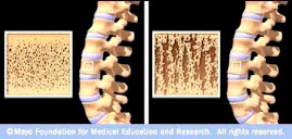
Source: www.mayoclinic.com