Micro computed tomography or "micro-CT" is x-ray imaging in 3D, by the same method used in hospital CT (or "CAT") scans, but on a small scale with massively increased resolution. It really represents 3D microscopy, where very fine scale internal structure of objects is imaged non-destructively.
“Postmenopausal osteoporosis is a chronic skeletal disorder involving compromised bone strength leading to an increased rate of fracture. Bone strength and therefore fracture risk depends on microarchitecture, matrix volume, and mineralization. Both cortical and trabecular bone are affected by the disease, with progressive considerable alterations in microarchitecture. The imbalance in bone remodeling in osteoporosis, in which bone formation is not able to compensate for bone resorption, leads to trabecular thinning and a loss of connectivity. As the disease progresses through years, continued increase remodeling rates lead to deep resorption cavities, the trabecular plates become perforated, and the connecting rods dissolve. As a consequence, there is a continuous shift from a plate‐like structure to a rod‐like structure. Simultaneously, in cortical bone, extensive remodeling at the endocortical surfaces “trabecularizes” (makes similar to trabecular bone) the inner part of cortical bone.”
“Cortical bone is the major component of long bone, for example, in the femoral shaft, in which rigidity to withstand load and movement is favored over flexibility. On the other hand, bone with a large proportion of trabecular bone is lighter and more flexible and can absorb more energy, for example, in the vertebrae. Both types of bone are susceptible to osteoporotic fracture at rates dependent on a number of variables, including microarchitecture.”
“The 3D analysis from the μCT studies goes beyond the histomorphometric results by showing differences on the trabecular and cortical microarchitecture of bone in favor of strontium ranelate compared with placebo (Fig. 1). In trabecular bone, Tb.N (trabecular number) was higher and Tb.Sp (trabecular separation) was lower in the treated group, indicating a thickening of the trabecular elements. The 22% reduction in trabecular SMI toward a value closer to zero means that 3‐yr treatment has shifted the structure of the cancellous bone away from a rod‐like structure (SMI = 3) toward a normal structure based on plates (SMI = 0). The significant improvement in two of these parameters with strontium ranelate is in line with an overall improvement in bone strength with this agent.”
“An increase in porosity or thinning in cortical bone reduces the resistance of bone to the propagation of cracks, which is one of the underlying defects in osteoporosis. Our 3D results showed that strontium ranelate significantly increases Ct.Th (cortical thickness) but had no effect on Ct.Po (cortical porosity). The anabolic agent teriparatide may actually increase Ct.Po.”

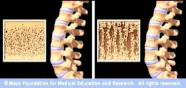
Figure 1. 3D microstructure from μCT of transiliac bone biopsies from postmenopausal osteoporotic
women receiving strontium ranelate (2 g/d) or placebo for 3 yr.