Human Skeleton

Showing posts with label dual-energy X-ray absorptiometry (DXA). Show all posts
Showing posts with label dual-energy X-ray absorptiometry (DXA). Show all posts
Monday, September 28, 2020


Friday, February 16, 2018
BMD Increases by Varying Amounts with Strontium Citrate and Decreases by Varying Amounts after Discontinuation
A study
published September 13, 2016, presents three case studies of patients who took
strontium citrate (680 mg strontium per day) for six years, five years, and
four years (Cases 1, 2, and 3, respectively) and then stopped taking it for two
years, one year, and one year (Cases 1, 2, and 3, respectively). The study
gives the increases in BMD with SrC and the decreases when the supplement was
discontinued. Of note is the fact that
both the increases and decreases vary with the site (lumbar spine or total hip),
the individual, and the number of years of use or discontinuation of SrC. Also,
keep in mind that stopping any of the osteoporosis drugs will also result in
loss of some of the BMD gains because there is no cure for osteoporosis.
Case 1
A 76-year-old
female with osteoporosis (OP) took alendronate weekly for 10 years till 2006,
then every other week for another year and was initiated on a drug holiday. She
started self-administrating SrC (680mg/day) in 2006 and continued to take daily
calcium (600mg bid) and vitamin D (2000units). Serum calcium and Vitamin D
(32-66ng/ml) remained normal. She continued SrC on her own till 2012, when she
agreed to stop taking it due to concerns about accumulation and toxicity.
On dual-energy X-ray absorptiometry (DXA) scan, total hip mean BMD increased 2.7% in
the first 2 years on SrC after stopping alendronate, and continued to increase
to a maximum of 9.2% after 6 years of treatment with SrC (1.53%/year). Lumbar
Spine (LS) BMD was non-diagnostic due to degenerative changes in the spine.
In the first 2 years off of SrC, total
hip mean BMD decreased by 5.7%.
Case 2
A 64-year-old
female with history of OP, which was diagnosed in 2001, took Actonel 35mg
weekly for 5 years, stopped for more than a year and resumed Actonel
intermittently for another 2 years before starting drug holiday. She was taking
Calcium 500 mg in the multivitamin and 2500 mg of vitamin D. Serum calcium and
vitamin D were within normal range. She started strontium citrate (680mg strontium)
daily in 2009 and took it for 5 years, stopping it in 2014.
Yearly
assessment of her BMD via DEXA scan showed an increase of 4.3% at LS spine
(L1-L4 vertebrae), and 7.6 % at total hip within 1 year of SrC treatment. At 5
years, BMD increase was 6.5 % at LS and 12% for mean total hip. She agreed to
stop SrC at this time.
One year after stopping SrC, LS BMD
decreased by 9.1 % and total hip BMD decreased by 4.2%.
Case 3
A 59-year-old female with OP was
treated with weekly alendronate for 4 years till 2007, when she stopped it due
to concerns for osteonecrosis of jaw. She was advised to resume alendronate in
2008 due to declining BMD, but opted not to take it.
She started self-administration of
680mg of SrC daily in February 2009 and continued it for four years till
January 2013, when she stopped it due to myalgias and concern about risk for
toxicity. Due to her history of ulcerative colitis, she was limited in dairy
intake, although she averaged half a cup of almond milk daily and took calcium
supplements. Serum calcium and vitamin D were within normal range.
BMD via DEXA scan showed an
increase of 10.7 % at LS (L1-L4) at 2 years after starting SrC, and 4.3 % at
total hip.
BMD
decreased by 14% at LS and by 6.4% at total hip a year after stopping SrC.
Mirza FS,
Azim S, Bhargava A (2016) Change in Bone Mineral Density with Strontium
Citrate: An Illusion or Reality. J Nutrition Health Food Sci 4(3): 1-3. DOI: http://dx.doi.org/10.15226/jnhfs.2016.00167
Monday, May 11, 2015
TBS iNsight® – a Novel Osteoporotic Fracture Assessment Diagnostic Tool
Medimaps Group
announced that a novel method for improving osteoporotic fracture
risk assessment, TBS iNsight®, is now globally available to
clinicians through a distribution partnership with General Electric Healthcare
(NYSE: GE). GE Healthcare provides transformational medical technologies and
services to meet the demand for increased access, enhanced quality and more
affordable healthcare around the world and is the market leader in the development
and manufacture of Dual Energy X-Ray Absorptiometry (DXA) bone densitometry
systems.
On June 10, 2014, Medimaps Group –
developers of the TBS iNsight® diagnostic tool – and General
Electric Healthcare signed a distribution agreement to distribute TBS
iNsight® on a global basis leveraging GE Healthcare’s extensive
direct sales and distribution organization.
TBS iNsight® is a unique,
easy-to-use software application that assesses bone texture - an index of bone
microarchitecture – which in turn is expressed as the Trabecular Bone Score
(TBS). The measurement is performed seamlessly using DXA images acquired from a
bone mineral densitometry (BMD) scan. No additional patient scan time or
radiation exposure is required and results are easily interpreted by physicians
to enable them to better manage patients with high risk of fractures. TBS
iNsight® has been cleared for marketing by the U.S. Food and Drug
Administration.
Prof. Didier Hans, Chairman and
Chief Executive Officer of Medimaps Group states; “It is well documented that
40-50% of osteoporotic fracture occurs in patients who are not determined to be
osteoporotic by BMD alone, yet BMD remains the only diagnostic method available
to most clinicians. A significant body of literature, including a recent study
of over 29,000 women, has demonstrated that TBS, when combined with hip and/or
spine BMD and FRAX® clinical factors, can help improve the
prediction of future osteoporotic fracture risk in a clinically meaningful way.
We’re very pleased to partner with General Electric Healthcare to bring this
valuable new predictive tool for better management of osteoporotic patients
across the globe.”
For more information on Medimaps and
TBS iNsight®, please visit www.medimapsgroup.com and www.tbsinsight.com
About Medimaps Group
Medimaps group – a Swiss company
founded by a group of clinical practitioners and researchers - is a developer
of innovative imaging software used in clinical practice for improved patient
management with minimum impact on the workflow of the practitioner. The company
has raised several rounds of venture capital to further commercialize TBS
iNsight® in the osteoporosis treatment market and for product
development of the TBS iNsight® tool for optimizing implant surgery
in the dental and orthopedic markets.
Contacts
Medimaps Group
Oscar Lazaro, +1 781-492-2012
Chief Commercial Officer
olazaro@medimapsgroup.com
Oscar Lazaro, +1 781-492-2012
Chief Commercial Officer
olazaro@medimapsgroup.com
Wednesday, May 6, 2015
Trabecular Bone Score
"The trabecular bone score (TBS) is a gray-level textural metric that can
be extracted from the two-dimensional lumbar spine dual-energy X-ray
absorptiometry (DXA) image. TBS is related to bone microarchitecture and
provides skeletal information that is not captured from the standard
bone mineral density (BMD) measurement. Based on experimental variograms
of the projected DXA image, TBS has the potential to discern
differences between DXA scans that show similar BMD measurements. An
elevated TBS value correlates with better skeletal microstructure; a low
TBS value correlates with weaker skeletal microstructure. Lumbar spine
TBS has been evaluated in cross-sectional and longitudinal studies."
"The following conclusions are based upon publications reviewed in this article:
1) TBS gives lower values in postmenopausal women and in men with previous fragility fractures than their nonfractured counterparts;
2) TBS is complementary to data available by lumbar spine DXA measurements;
3) TBS results are lower in women who have sustained a fragility fracture but in whom DXA does not indicate osteoporosis or even osteopenia;
4) TBS predicts fracture risk as well as lumbar spine BMD measurements in postmenopausal women;
5) efficacious therapies for osteoporosis differ in the extent to which they influence the TBS;
6) TBS is associated with fracture risk in individuals with conditions related to reduced bone mass or bone quality."
"Based on these data, lumbar spine TBS holds promise as an emerging technology that could well become a valuable clinical tool in the diagnosis of osteoporosis and in fracture risk assessment."
http://www.ncbi.nlm.nih.gov/pubmed/24443324
"The following conclusions are based upon publications reviewed in this article:
1) TBS gives lower values in postmenopausal women and in men with previous fragility fractures than their nonfractured counterparts;
2) TBS is complementary to data available by lumbar spine DXA measurements;
3) TBS results are lower in women who have sustained a fragility fracture but in whom DXA does not indicate osteoporosis or even osteopenia;
4) TBS predicts fracture risk as well as lumbar spine BMD measurements in postmenopausal women;
5) efficacious therapies for osteoporosis differ in the extent to which they influence the TBS;
6) TBS is associated with fracture risk in individuals with conditions related to reduced bone mass or bone quality."
"Based on these data, lumbar spine TBS holds promise as an emerging technology that could well become a valuable clinical tool in the diagnosis of osteoporosis and in fracture risk assessment."
http://www.ncbi.nlm.nih.gov/pubmed/24443324
Thursday, December 18, 2014
Study Demonstrates DXA Scan Is Obviously Flawed
Most clinically important vertebral
body compression fractures found on abdominal multidetector computed tomography
(CT) scans would go unreported if sagittal reconstructions were not routinely
evaluated, according to a new study.
"At this point, it is routine
to evaluate sagittal reconstructions at abdominal multidetector CT (MDCT), and
whenever vertebral fractures are detected, they are reported." However,
this has not always been the case, said Travis Lauder, BS, a fourth-year
medical student at the University of Wisconsin School of Medicine and Public
Health in Madison.
"The studies we reviewed were
done before 2007, when sagittal reconstructions were not automatically
provided. We showed that without them, we missed a lot of vertebral compression
fractures in patients who underwent abdominal multidetector CT," he
explained.
Lauder presented the study results
at the American Roentgen Ray Society 2013 Annual Meeting in Washington, DC.
First results were published online February 28 in Radiology.
Lauder and his team retrospectively
analyzed 2041 consecutive patients (mean age, 70.8 years) who had undergone
abdominal multidetector CT and dual-energy x-ray absorptiometry (DXA) within 6
months of each other. The images were taken from 2000 to 2007, before sagittal
CT reconstructions were routinely obtained.
The researchers used the Genant
visual semiquantitative method to review transverse (axial) and retrospective
sagittal multidetector CT reconstructions for the presence of moderate to
severe fractures of the lower thoracic and lumbar spine. They also reviewed the
electronic medical records of these patients to determine whether the fracture
was reported at prospective CT interpretation, was known previously, or was
subsequently diagnosed.
"At least 1 moderate or severe
vertebral body compression fracture was retrospectively identified on CT in 97
patients," the researchers report. The fractures involved 1 level in 67
patients and 2 or more levels in 30 patients. In total, 141 fractures were
identified.
In patients with an unreported fracture, vertebral body
compression fracture was not recognized clinically in 64% and there was no
evidence of osteoporosis on DXA in 48%.
"DXA is still the gold
standard," Lauder noted, "but
this analysis demonstrated that a lot of these patients have false negatives on
DXA, and that the DXA test is obviously flawed."
Incidental findings are a well-known
"double-edged sword" in cross-sectional imaging, said Abraham
Dachman, MD, professor of radiology and director of the fellowship programs at
the University of Chicago in Illinois, who was asked by Medscape Medical
News to comment on the study.
"Many findings — some
previously known by the referring physician and some not — can be found on CT.
It is often difficult for the radiologist to know what findings to report, what
findings to ignore, and what incidental findings are sufficiently important to
require personal communication with the referring physician," Dr. Dachman
said.
However, this study highlights the
potential of abdominal CT to detect vertebral body compression fractures, he
noted. Coronal and sagittal reconstructions are often done by the technologist
who performs the exam using software on the CT scanner console; they do not
require dedicated workstations.
"This study did not address how
often the observation of vertebral body compression fractures changed patient
management because it was retrospective in nature," Dr. Dachman said.
"Still, it highlights the potential of CT to reveal information and
provides a rationale to radiologists who wish to routinely report this
observation."
This article appeared on Medscape
under the title, “Vertebral Fractures Go Undetected without Sagittal Views.”
The abstract and full text with CT
images for “Unreported Vertebral Body Compression Fractures at Abdominal Multidector
CT,” which was published in Radiology
in July 2013, can be read here:
For an excellent description of computed
tomography (CT) and multidetector computed tomography (MDCT), see:
For a description of an abdominal CT
scan, see:
For information on computed tomography
angiography (CTA), see:
Subscribe to:
Posts (Atom)
Wandering Skeleton

Artist: Joel Hoekstra
Osteoporotic Bone
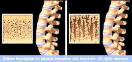
Source: www.mayoclinic.com