The potential
effect of strontium (Sr) on bone apatite (bone mineral) crystals was
investigated in paired biopsies of osteoporotic women treated with either
strontium ranelate (SrRan) or a placebo for 36 months. In ten paired biopsies,
crystallinity, apparent length and width/thickness of crystals, interplanar
distances, and lattice parameters of unit cells were assessed by X-ray
diffraction and selected area electron diffraction. None of these parameters,
reflecting crystal and unit cell characteristics, was influenced by the
presence of Sr. All of the parameters were similar in SrRan and placebo groups
after 36 months of treatment. The mean rate of substitutions of calcium by Sr
ions was 4.5 %. Overall, the quality of bone apatite crystals was maintained
after 36 months of treatment with SrRan. Results of this study were published on
March, 2013, in Osteoporosis
International.
http://www.ncbi.nlm.nih.gov/pubmed/23108780Human Skeleton

Monday, October 21, 2013


Friday, September 13, 2013
Protein Believed to Cause Postmenopausal Osteoporosis
Researchers have
identified a protein that likely causes osteoporosis in women after menopause. In
experiments on mice, the research team from Tokyo’s Keio University School of
Medicine and other institutions found that a decrease in the secretion of
estrogen enhances the function of HIF1 alpha protein. Once the amount of
estrogen secreted by the ovaries begins to decline around the time of
menopause, HIF1 alpha protein in osteoclasts increases.
Normal mice after menopause showed decreased femoral bone density compared with premenopausal levels, but mice whose genes were manipulated so as not to produce the HIF1 alpha protein did not show a decline of femoral bone density even after menopause. Moreover, a substance to inhibit the function of the HIF1 alpha protein that was given to postmenopausal mice led to an increase of bone density compared with premenopausal levels. The discovery is expected to lead to the development of an effective medicine to prevent osteoporosis.
The study is to be published in the online edition of the Proceedings of the U.S. National Academy of Science.
Normal mice after menopause showed decreased femoral bone density compared with premenopausal levels, but mice whose genes were manipulated so as not to produce the HIF1 alpha protein did not show a decline of femoral bone density even after menopause. Moreover, a substance to inhibit the function of the HIF1 alpha protein that was given to postmenopausal mice led to an increase of bone density compared with premenopausal levels. The discovery is expected to lead to the development of an effective medicine to prevent osteoporosis.
The study is to be published in the online edition of the Proceedings of the U.S. National Academy of Science.
Sunday, September 1, 2013
From Osteoporosis to Osteopenia with Strontium Citrate
After five and a half years of taking strontium citrate (680
mg strontium daily), I went from osteoporosis to osteopenia. I was probably at
the osteopenia stage much earlier, but I waited four years between my previous
DEXA scan, which showed improvement at all sites but still left me with
osteoporosis, and this one. My density increased 34% in the lumbar spine and
55% in the left hip! These numbers are phenomenal! Even if up to 50% of the
change in BMD were attributed to the effect of strontium, which has a higher
atomic number than calcium and will affect DEXA measurements, the numbers would
still be great. My BMD is now 0.871 g/cm2 at the spine from L1- L4 (T-score -1.6),
0.692 g/cm2 at the left femoral neck (-1.4 T-score), and 0.775 g/cm2 at the
left total hip (-1.4 T-score). Osteopenia is defined as T-scores < -1.0 and
-2.5.
Monday, August 26, 2013
Non-Invasive Measurement of Bone Strontium Levels
Many of you have probably read that
the only way to measure the amount of strontium in bone is to do a bone biopsy.
That is no longer true, at least not in a research setting. A case study
published last year in Bone used an in vivo X-ray
fluorescence (IVXRF) I-125 based system to measure bone strontium levels
non-invasively in an osteoporotic female volunteer before and after she began
taking strontium citrate supplements (680 mg Sr/day).
Thirty-minute
measurements were taken at the finger and ankle bone sites, representing
primarily cortical and trabecular bone, respectively. Baseline natural strontium levels were obtained followed by a 24h measurement of first
intake of strontium citrate supplements (680 mg Sr/day). The baseline levels of
strontium (prior to supplementation) were 0.38 ± 0.05 and 0.39 ± 0.10 for the
finger and ankle, respectively. After 24 hrs the levels were 0.62 ± 0.14 and
0.45 ± 0.12 for the finger and ankle, respectively. By 120 h, the increase was
statistically significant at 0.68 ± 0.07 and 0.93 ± 0.05, respectively. Further
increases occurred within an interval of 90-180 days, with the most recent,
after 800 days, at the finger and ankle being 7 and 15 times higher than the
initial baseline reading. The results show bone strontium incorporation and retention follow a pattern and suggest strontium levels, at least in the ankle, do not plateau within two to three years and will continue to increase over time, as an individual takes strontium supplements. The ability of this IVXRF system to monitor and measure bone strontium levels over time provides a useful diagnostic tool to help gain insight into strontium bone kinetics.
Monday, August 5, 2013
Healing Fractures with Strontium Ranelate
Age and bone
quality are the two most important factors influencing the fracture healing
process. Cellular and molecular alterations in elderly patients may lead to pseudarthrosis
(nonunion), i.e., a fracture that has not united in the stipulated time in
which such fractures usually unite and has no chance of union without
intervention. In the same way, in altered bone metabolism conditions, such as
osteoporosis, physiological phases of fracture healing are impaired.
Two women with
fractures that would not heal were started on strontium ranelate (2g/day),
calcium (1200 mg/day), and vitamin D (800 IU/day). Patient B.G. was a
57-year-old Caucasian woman with osteopenia and a fracture of the right distal
radius and ulna. Patient S.L. was a 59-year-old Caucasian woman with
osteoporosis and a fracture of the base of the fifth metatarsal in the left
foot. Their case studies showed healing of fractures in a short period of time
after drug and supplement intervention.
Read more about
these fascinating studies, which contain figures of several radiographs and a
CT scan, at http://www.ncbi.nlm.nih.gov/pmc/articles/PMC2898010/
Monday, April 29, 2013
EMA Confirms Recommendations to Restrict Strontium Ranelate
The European Medicines Agency (EMA) has confirmed the recommendations to restrict the use of strontium ranelate (Protelos/Osseor, Servier) due to concerns about the risk of adverse
cardiac events. The EMA's Pharmacovigilance Risk Assessment
Committee (PRAC) recommended the restrictions earlier this month, and the
Committee for Medicinal Products for Human Use (CHMP) has now endorsed these following its meeting of April 22-25, 2013. The
CHMP opinion will be sent to the European Commission, the executive branch of
the European Union, which will issue a legally binding decision.
Subscribe to:
Posts (Atom)
Wandering Skeleton

Artist: Joel Hoekstra
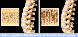
Osteoporotic Bone
Source: www.mayoclinic.com