Human Skeleton

Human Skeleton

WELCOME TO STRONTIUM FOR BONES BLOG

Have you experienced negative, and even dangerous, side effects from Fosamax (alendronate), Boniva (ibandronate), Actonel (risedronate), Reclast (zoledronic acid), Prolia (denosumab), Forteo (teriparatide), Tymlos (abaloparatide), or other drugs prescribed for osteoporosis? If you have, then rest assured there is a safe, effective treatment for this condition. Strontium, primarily in the form of strontium citrate, is taken orally once a day.

Visitors to my blog can leave comments or ask questions and can remain anonymous, if they wish. Their comments are relayed to my g-mail inbox. Below each post, the number of comments for that post is cited and underlined because it is a link. By clicking on that link below any post, a window opens so that a visitor can leave a comment. Ideally, visitors leave comments on posts most relevant to their comments. All comments to my posts are moderated by me.

Browse the posts and visit the link library of references.


Blog Archive

Friday, March 13, 2015

Women's Mid-Life Non-Traumatic Fracture Risk Higher With Low Vitamin D


The lower a woman's vitamin D levels as she transitions to menopause, the greater her risk of sustaining a non-traumatic fracture. 

"Women who have levels less than 20 nanograms per milliliter would need some type of vitamin D supplementation," Dr. Jane A. Cauley of the University of Pittsburgh, who worked on the study, told Reuters Health. The findings back up the Institute of Medicine's conclusion that levels lower than 20 ng/ml are insufficient for skeletal health, she added.

Bone loss is known to accelerate during the menopausal transition, and postmenopausal women require significantly more calcium to reach neutral calcium balance than perimenopausal women, Dr. Cauley and her team note in their report, online February 23 in the Journal of Clinical Endocrinology and Metabolism.

To determine if serum 25(OH)D levels would be associated with changes in bone mineral density (BMD) and fracture risk, the researchers looked at data from the Study of Women's Health Across the Nation (SWAN) on 124 women with an incident traumatic fracture, 88 with an incident non-traumatic fracture, and 1,532 who did not sustain fractures during the study's average follow-up period of 9.5 years. 

Each woman had her serum 25(OH)D level checked at her third annual visit, while her BMD was assessed annually. The women's mean age at the study's outset was 48.5.

The study participants' mean 25(OH)D levels were 21.8 ng/ml, and 43% had levels below 20 ng/mL. While multivariate analysis found no association between 25(OH)D levels and traumatic fracture risk, the risk of non-traumatic fractures decreased by 28% for every 10 ng/ml increase in serum 25(OH)D. [Referring to Table 3 of the original article, “For every 10 ng/ml increase in 25(OH)D, the hazard ratio (HR) for nontraumatic fracture was 0.72 (0.54, 0.95).”; 1.00 minus 0.72 equals 0.28, or 28%]

Women with levels of 20 ng/ml or higher had a 46% lower risk of fracture during follow-up than women with lower levels of 25(OH)D. However, there was no association between baseline 25(OH)D and changes in BMD at the lumbar spine or femoral neck across menopause.

Roughly 22% of the women in the study had vitamin D levels greater than 30 ng/ml, Dr. Cauley said, so the study lacked power to determine if these women's non-traumatic fracture risk was further reduced.
"We didn't see any relationship between 25(OH)D and these bone changes, but we were somewhat limited in that we didn't have any information on how vitamin D changed across menopause," she added.


http://www.nutrition411.com/news/womens-mid-life-non-traumatic-fracture-risk-higher-low-vitamin-d
http://www.medscape.com/viewarticle/841028?src=wnl_edit_tpal&uac=127701PY:
http://press.endocrine.org/doi/pdf/10.1210/jc.2014-4367

Thursday, March 12, 2015

Strontium Prevents the Progression of Thoracic Kyphosis


Background: Thoracic kyphosis (a frequent feature in the elderly and in post menopausal osteoporosis) can be caused by vertebral fractures. This exaggerated curvature of the spine is also related to degenerative changes including intervertebral disc space narrowing, deformities of the anterior part of the vertebrae and reduced spinal muscles strength. An increase over time in thoracic kyphosis has been associated with impairment in global health due to increased body sway and risk of falls, impairments in pulmonary function, presence of esophagial hiatal hernia, and an increase in risk of mortality in older women.

Objectives: The objective of this study was to assess the effect on thoracic kyphosis progression over a 3-year treatment with strontium ranelate, a treatment against osteoporosis that reduces the risk of vertebral, nonvertebral and hip fractures.

Methods: This study was performed in women with postmenopausal osteoporosis from SOTI 1 and TROPOS 2 studies, aiming to demonstrate the efficacy of strontium ranelate against vertebral and non-vertebral fractures. Patients underwent lateral radiographs of the thoracic and lumbar spine at baseline and annually over 3 years (standardized procedures). The level of thoracic kyphosis was reflected by a kyphosis index, defined on lateral thoracic radiographs as the ratio BD/AC (AC= line from the anterior superior edge of T4 to the anterior inferior edge of T12; BD= perpendicular line from the furthest superior or inferior posterior point of T7, T8 or T9 vertebrae to AC line) expressed as a %. The highest the KI, the more severe the thoracic kyphotic curvature.

Results: The population consisted of 4055 women with postmenopausal osteoporosis (2038 patients randomised to strontium ranelate and 2017 randomised to placebo). Baseline characteristics were similar: mean age 73.5; Spine BMD T-score (L2-L4): -3.06; Femoral neck T-score: -2.97; KI: 25.4. Over 3 years, there was a significant increase from baseline in kyphotic index for all patients. However, this increase was significantly lower (p=0.003) in patients treated with strontium ranelate: +3.71±7.69% than in the placebo group:+4.70±7.32%.
The same calculations were repeated after exclusion of patients having either prevalent or incident thoracic vertebral fractures. In this subset of 1193 patients (634 in the strontium ranelate group and 559 in the placebo group), the change in the strontium ranelate group was still lower than in the placebo group (+ 2.72±7.17% versus 4.34±6.54%, respectively).


Conclusion: These prospective results demonstrate that thoracic kyphosis increases over time in postmenopausal women with osteoporosis. Strontium ranelate decreased the progression of this thoracic kyphosis over 3 years, regardless of the presence or not of vertebral fractures. This new effect of strontium ranelate possibly reflects additional benefit on spinal components besides its efficacy in decreasing vertebral fractures.

References: 1 Meunier PJ et al. N Engl J Med 2004; 2 Reginster J.Y et al. JCEM 2005

Ann Rheum Dis 2008;67(Suppl II):541
http://www.abstracts2view.com/eular/view.php?nu=EULAR08L_SAT0344

Friday, March 6, 2015

Too Much Vitamin D Can Be Bad for You


Context:
Optimal levels of vitamin D have been a topic of heavy debate, and the correlation between 25-hydroxyvitamin D [25(OH)D] levels and mortality still remains to be established.

Objective:
The aim of the study was to determine the association between all-cause mortality and serum levels of 25(OH)D, calcium, and PTH.

Design and Setting:
We conducted a retrospective, observational cohort study, the CopD Study, in a single laboratory center in Copenhagen, Denmark.

Participants:
Serum 25(OH)D was analyzed from 247,574 subjects from the Copenhagen general practice sector. In addition, serum levels of calcium, albumin-adjusted calcium, PTH, and creatinine were measured in 111,536; 20,512; 34,996; and 189,496 of the subjects, respectively.

Main Outcome Measures:
Multivariate Cox regression analysis was used to compute hazard ratios for all-cause mortality.

Results:
During follow-up (median, 3.07 yr), 15,198 (6.1%) subjects died. A reverse J-shaped association between serum level of 25(OH)D and mortality was observed. A serum 25(OH)D level of 50–60 nmol/liter was associated with the lowest mortality risk. Compared to 50 nmol/liter, the hazard ratios (95% confidence intervals) of all-cause mortality at very low (10 nmol/liter) and high (140 nmol/liter) serum levels of 25(OH)D were 2.13 (2.02–2.24) and 1.42 (1.31–1.53), respectively. Similarly, both high and low levels of albumin-adjusted serum calcium and serum PTH were associated with an increased mortality, and secondary hyperparathyroidism was associated with higher mortality (P < 0.0001).

Conclusion:
In this study from the general practice sector, a reverse J-shaped relation between the serum level of 25(OH)D and all-cause mortality was observed, indicating not only a lower limit but also an upper limit. The lowest mortality risk was at 50–60 nmol/liter. The study did not allow inference of causality, and further studies are needed to elucidate a possible causal relationship between 25(OH)D levels, especially higher levels, and mortality.

Note: 50-60 nmol/L equals 20-24 ng/ml. To convert nmol/L to ng/ml, divide by 2.5. To convert ng/ml to nmol/L, multiply by 2.5.  

The full article is available here:


Wednesday, March 4, 2015

Exercise Rapidly Improves Bodies and Brains


Identical twins in Finland who shared the same sports and other physical activities as youngsters but different exercise habits as adults soon developed quite different bodies and brains, according to a new study that highlights the extent to which exercise shapes our health, even in people who have identical genes and nurturing.

The researchers found 10 pairs of young, male identical twins, one of whom regularly exercised, while the other did not. The dissimilarities in their exercise routines had mostly begun within the past three years, according to their questionnaires.

The scientists measured each man’s endurance capacity, body composition and insulin sensitivity, to determine their fitness and metabolic health. The scientists also scanned each twin’s brain.

The sedentary twins had lower endurance capacities, higher body fat percentages, and signs of insulin resistance, signaling the onset of metabolic problems. (The twins tended to have similar diets, so food choices were unlikely to have contributed to health differences.)

The twins’ brains also were unalike. The active twins had significantly more gray matter than the sedentary twins, especially in areas of the brain involved in motor control and coordination.

The researchers concluded the following:
“Among healthy adult male twins in their mid-30s, a greater level of physical activity is associated with improved glucose homeostasis and modulation of striatum and prefrontal cortex gray matter volume, independent of genetic background. The findings may contribute to later reduced risk of type 2 diabetes and mobility limitations.”

The study was small and not a formal randomized trial. But the results strongly imply that the differences in the twin’s exercise habits caused the differences in their bodies.

More subtly, the findings also point out that genetics and environment do not have to be destiny when it comes to exercise habits. Even if the input from our DNA and upbringing urges us to skip the gym, we can move more, and, based on this study, rapidly and substantially improve the condition of our bodies and brains.


Tuesday, January 27, 2015

Fracture Risk


“Osteoporosis and resultant fractures of the spine, hip and other sites are important public health problems with significant individual and societal costs. The risk for osteoporotic fracture is based upon low bone density and the presence of one or more clinical risk factors (see Table 1). A history of fracture during adulthood or falls are important clinical factors in determining the risk of future fracture; however, age is the most influential risk factor, such that middle-aged adults with other risk factors are likely to be at low absolute fracture risk in the medium term. Using these clinical risk factors and BMD when available, fracture risk assessment tools (based upon data collected from large prospective observational studies) have been developed to estimate the 5–10 year probability of hip fracture and other fractures in untreated patients. Clinicians should be aware that fracture risk can also be estimated using the FRAX or Garvan tools without BMD data. Chronic glucocorticoid use is an established risk factor for osteoporosis, with studies showing that use of glucocorticoids leads to accelerated bone loss and an increased risk of fracture. Other drugs are increasingly recognized as potential causes of bone loss and fracture, particularly amongst predisposed individuals….”

See the following link for more on other drugs that can cause bone loss and fractures:

Table 1.  Clinical risk factors for fracture
Advancing age
Previous fracture during adulthood
History of a fall or falls in the past 12 months
Glucocorticoid therapy
Parental history of hip fracture
Low body weight
Current cigarette smoking
Excessive alcohol consumption
Medical diseases (e.g. rheumatoid arthritis, hyperparathyroidism, coeliac disease, hypogonadism)

This introduction and table appeared in “Adverse Skeletal Effects of Drugs – Beyond Glucocorticoids,” Susannah O'Sullivan, Andrew Grey, Clin Endocrinol. 2015; 82(1):12-22. 
http://www.medscape.com/viewarticle/837369?src=wnl_edit_tpal&uac=127701PY

Wandering Skeleton

Wandering Skeleton
Artist: Joel Hoekstra

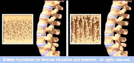
Osteoporotic Bone

Osteoporotic Bone
Source: www.mayoclinic.com

How Strontium Builds Bones

Strontium is a mineral that tends to accumulate in bone. Studies have shown that oral doses of strontium are a safe and effective way to prevent and reverse osteoporosis. Doses of 680 mg per day appear to be optimal. See my "For More Information About Strontium" links section.

Osteoporosis is caused by changes in bone production. In healthy young bones there is a constant cycle of new bone growth and bone removal. With age, more bone is removed and less new bone is produced. The bones become less dense and thus more fragile.

Scientists believe that strontium works in two ways. It may stimulate the replication of pre-osteoblasts, leading to an increase in osteoblasts (cells that build bone). Strontium also directly inhibits the activity of osteoclasts (cells that break down bone). The result is stronger bones.

When taking strontium, be sure to take 1200 mg calcium, 1000 IU vitamin D3, and 500 mg magnesium daily. It is best to take strontium late at night on an empty stomach. Calcium and strontium may compete with each other for absorption if taken together.