I will be asking that a Trabecular Bone Score (TBS) be included with my next DXA scan. The doctor who read my latest scan wrote, "Follow up with DEXA and TBS: As needed."
I found a review of TBS. The review includes a section on "Changes in TBS with Treatment of Osteoporosis." Below is the paragraph comparing the effects of strontium ranelate and alendronate (Fosamax) on TBS.
"The effects of strontium ranelate (SrRan) and alendronate on TBS were evaluated in a post hoc analysis performed in 79 women with postmenopausal osteoporosis of 189 included in a double‐blind, double‐dummy, randomized study. Women were randomized to either SrRan 2 g/day or alendronate 70 mg/week for 2 years. TBS and BMD parameters were assessed in the LS after 12 and 24 months of treatment. Over 1 and 2 years, LS BMD increased significantly by 5.6% and 9.0% in the SrRan group and by 5.2% and 7.6%, respectively, in the alendronate group. LS TBS increased by 2.3% (p < 0.001) and 3.1% (p < 0.001) in the SrRan group, but the change in the alendronate group was not significant (0.5% and 1.0%, respectively). There was a significant between‐group difference with SrRan showing larger TBS increases than alendronate."
Let me reiterate: Over one and two years, Lumbar Spine (LS) BMD increased in both the SrRan (5.6%, 9.0%) and alendronate groups (5.2%, 7.6%). You will note that the SrRan BMD numbers are higher, especially after the second year, than the alendronate numbers. The results are as expected because strontium results in an overestimation of BMD.
HERE IS THE KICKER: LS TBS increased by 2.3% and 3.1% in the SrRan group, but the change in the alendronate group was not significant (0.5% and 1.0%, respectively). There was a significant between‐group difference with SrRan showing larger TBS increases than alendronate.
Keep in mind that TBS is related to bone microarchitecture and provides skeletal information that is not captured from the standard BMD measurement. TBS may be a better predictor of fracture risk than BMD alone.
https://www.panoramaortho.com/wp-content/uploads/2019/03/TBS-Rev...
Human Skeleton

Showing posts with label fracture risk. Show all posts
Showing posts with label fracture risk. Show all posts
Tuesday, August 23, 2022
Trabecular Bone Score (TBS)


Sunday, July 5, 2015
B Vitamins and Homocysteine and Fracture Risk
“Some studies suggest high blood
levels of the amino acid homocysteine may be linked to lower bone density and
higher risk of hip fracture in the elderly. Vitamins B6 and B12, as well as
folic acid, play a role in changing homocysteine into other amino acids for use
by the body, so it is possible that they might play a protective role in
osteoporosis. Research is ongoing as to whether supplementation with these B
vitamins might reduce fracture risk4,5.”
4. McLean RR, Jacques PF, Selhub J, et al. (2004)
Homocysteine as a predictive factor for hip fracture in older persons. N Engl J
Med 350:2042-49
5. Morris MS, Jacques PF, Selhub J (2005) Relation between homocysteine and
B-vitamin status indicators and bone mineral density in older Americans. Bone
37:234-42
WebMD ranks the effectiveness of vitamin B12 for various
uses. The rankings are “effective for,” “likely effective for,” “possibly
effective for,” “possibly ineffective for,” and “insufficient evidence for.”
According to webMD, vitamin B12 is likely effective for “high
level of homocysteine in the blood (Hyperhomocysteinemia).” “Taking vitamin B12
by mouth, along with folic acid and sometimes pyridoxine (vitamin B6), can
lower blood levels of homocysteine.”
http://www.webmd.com/vitamins-supplements/ingredientmono-926-vitamin%20b12.aspx?activeingredientid=926&activeingredientname=vitamin%20b12
Tuesday, January 27, 2015
Fracture Risk
“Osteoporosis and resultant fractures of the spine, hip and
other sites are important public health problems with significant individual
and societal costs. The risk for osteoporotic fracture is based upon low bone
density and the presence of one or more clinical risk factors (see Table 1). A
history of fracture during adulthood or falls are important clinical factors in
determining the risk of future fracture; however, age is the most influential
risk factor, such that middle-aged adults with other risk factors are likely to
be at low absolute fracture risk in the medium term. Using these clinical risk
factors and BMD when available, fracture risk assessment tools (based upon data
collected from large prospective observational studies) have been developed to
estimate the 5–10 year probability of hip fracture and other fractures in
untreated patients. Clinicians should be aware that fracture risk can also be
estimated using the FRAX or Garvan tools without BMD data. Chronic
glucocorticoid use is an established risk factor for osteoporosis, with studies
showing that use of glucocorticoids leads to accelerated bone loss and an
increased risk of fracture. Other drugs are increasingly recognized as
potential causes of bone loss and fracture, particularly amongst predisposed
individuals….”
See the following link for more on other drugs that can
cause bone loss and fractures:
|
Table
1. Clinical risk factors for fracture
|
|
Advancing age
|
|
Previous fracture during adulthood
|
|
History of a fall or falls in the
past 12 months
|
|
Glucocorticoid therapy
|
|
Parental history of hip fracture
|
|
Low body weight
|
|
Current cigarette smoking
|
|
Excessive alcohol consumption
|
|
Medical diseases (e.g. rheumatoid
arthritis, hyperparathyroidism, coeliac disease, hypogonadism)
|
This introduction and table
appeared in “Adverse Skeletal Effects of Drugs – Beyond Glucocorticoids,” Susannah O'Sullivan, Andrew Grey, Clin
Endocrinol. 2015; 82(1):12-22.
http://www.medscape.com/viewarticle/837369?src=wnl_edit_tpal&uac=127701PY
Monday, January 26, 2015
Adverse Skeletal Effects of Drugs
There
is an expanding list of drugs for which there are concerns regarding adverse
skeletal effects. The effect of long-term use of glucocorticoid drugs to cause
accelerated bone loss and increase the risk of fracture is well recognized. For
some drugs (TKIs, calcineurin inhibitors and loop diuretics), the available
data are inconsistent and/or do not support a definite adverse skeletal effect.
Patients receiving these drugs can be managed in line with guidelines for the
general population (See table). Other drugs (SSRIs, antipsychotic drugs, AEDs
and PPIs) do not seem to affect bone metabolism or BMD, but there is evidence
for increased fracture risk. However, the evidence is limited to observational
data, and any relationship may be attributable either to nonbone effects of the
drug or effects of the underlying condition to increase fracture rate. In these
patients, evaluation for other risk factors for fracture and management of
these risk factors should be considered. In a third group (depot MPA,
chemotherapy and ART), there is evidence for increased bone loss and/or
fracture, but the patient population is generally at low risk for fracture. In
these cases, patients may require an assessment of risk, but will rarely
require specific treatment for fracture risk reduction. In the last group
(aromatase inhibitors, GnRH agonists and thiazolidinediones), there is evidence
of increased risk of bone loss and/or fracture, and the drugs are more
frequently prescribed to individuals at higher baseline fracture risk. Patients
receiving these drugs require risk assessment and those at high risk of
fracture should receive alternative treatments (or "add-back" HRT in
the case of GnRH agonists) or, if necessary, specific treatment for
osteoporosis.
Table:
Summary of management recommendations
|
||
Example drugs
|
Evidence for risk
|
Management recommendations
|
Tyrosine kinase inhibitors
Calcineurin inhibitors Loop diuretics |
No adverse
skeletal effect or data inconsistent |
Manage as per general population
|
Selective serotonin re-uptake
inhibitors (SSRIs)
Antipsychotics Anti-epileptic drugs Proton pump inhibitors |
Evidence for increased fracture
risk but fracture risk seems to relate to nonbone effects of the drug or
effects of the underlying condition
|
Evaluate for other risk factors
for fracture and manage these risk factors
|
Depot medroxyprogesterone acetate
Chemotherapy Antiretroviral therapy |
Evidence for increased bone
loss and/or fracture, but low risk population |
Assessment of fracture risk, but
will rarely require specific treatment
|
Aromatase inhibitors
Gonadotrophin hormone-releasing hormone agonists Thiazolidinediones |
Evidence for increased risk of
bone loss and/or fracture, and the drugs are more frequently prescribed to
individuals at higher baseline fracture risk
|
Assessment of fracture risk and
those at high risk should receive alternative treatments and/or specific
treatment for osteoporosis.
|
The
conclusions and table are from a longer article, “Adverse Skeletal Effects of
Drugs – Beyond Glucocorticoids,” by Susannah O'Sullivan and Andrew Grey, Clin
Endocrinol. 2015;82(1):12-22.
Monday, August 11, 2014
Use of Computed Tomography for Assessing Bone Mineral Density

Dual x-ray absorptiometry (DXA) is
currently the standard for assessing bone mineral density (BMD) and has been
correlated with fracture risk and treatment efficacy. DXA includes the
posterior elements of the spine, and therefore
may be inaccurate or not possible in cases of severe spinal degeneration,
scoliosis, or following lumbar surgery. T-score evaluations are somewhat
limited in clinical utility, as the majority of patients who sustain fragility
fractures are not in the osteoporotic range.
Assessing local bone quality on CT scans with Hounsfield
unit (HU) quantification is being used with increasing frequency. Correlations
between HU and bone mineral density have been established, and normative data
have been defined throughout the spine. Recent investigations have explored the
utility of HU values in assessing fracture risk, implant stability, and spinal
fusion success. The information provided by a simple HU measurement can alert
the treating physician to decreased bone quality, which can be useful in both
medically and surgically managing these patients.
The purpose of this paper is to
review the reliability and validity of the techniques used to estimate bone
health using CT scans with Hounsfield unit (HU) quantification. Such scans can
be used to identify patients at risk for osteoporosis, and these values could
be used for surgical planning in cases of trauma, degeneration, and deformity.
There was good correlation of HU value to DXA for both BMD and T-score.
Subscribe to:
Posts (Atom)
Wandering Skeleton
Artist: Joel Hoekstra
Osteoporotic Bone
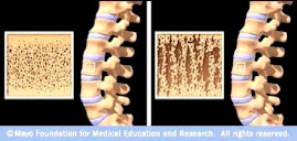
Source: www.mayoclinic.com