Human Skeleton

Human Skeleton

WELCOME TO STRONTIUM FOR BONES BLOG

Have you experienced negative, and even dangerous, side effects from Fosamax (alendronate), Boniva (ibandronate), Actonel (risedronate), Reclast (zoledronic acid), Prolia (denosumab), Forteo (teriparatide), Tymlos (abaloparatide), or other drugs prescribed for osteoporosis? If you have, then rest assured there is a safe, effective treatment for this condition. Strontium, primarily in the form of strontium citrate, is taken orally once a day.

Visitors to my blog can leave comments or ask questions and can remain anonymous, if they wish. Their comments are relayed to my g-mail inbox. Below each post, the number of comments for that post is cited and underlined because it is a link. By clicking on that link below any post, a window opens so that a visitor can leave a comment. Ideally, visitors leave comments on posts most relevant to their comments. All comments to my posts are moderated by me.

Browse the posts and visit the link library of references.


Blog Archive

Showing posts with label femoral neck. Show all posts

Wednesday, January 8, 2020

Why Do Hips Fracture?

"Normal healthy bones can be thought of as nature's scaffold poles. The tightly packed minerals that make up the cortical bone form a sheath around an inner core of spongy bone and provide the strength that supports our bodies. Throughout our lives, our skeletons are kept strong by the continuous creation of new, fresh bone and the destruction of old, worn out bone. Unfortunately, as we become older, destruction becomes faster than creation, and so the cortical layer thins, causing the bone to weaken and break more easily. In severe cases, this is known as osteoporosis. As a result, simple trips or falls that would only bruise a younger person can cause serious fractures in the elderly. However, half of the elderly patients admitted to hospital with a broken hip do not suffer from osteoporosis.

So why do those hips break? This is an important question because hip fractures are very debilitating. Repairing a fractured hip requires traumatic surgery that, even if successful, may not enable a patient to regain the full mobility they had beforehand. The National Osteoporosis Society estimates that 13,800 people in the UK die every year as a direct result of hip fractures. This is over 10% of patients injured (NHS Choices). Therefore, understanding how these fractures occur and acting to prevent them is vital for improving the quality and length of life of our aging population.

To better understand why hips fracture, researchers in Cambridge and Prague analysed CT scans performed on the opposite, unbroken hips of a group of elderly women admitted to Bulovka University Hospital in Prague with hip fractures (Poole et al., 2012). Previous studies have shown that these unbroken hips tend to be in a similar condition to the patient's other hip before it was fractured.

CT scanners are now standard pieces of equipment in most hospitals, and are used to examine organs and tissues inside the body. Essentially a rotating X-ray machine, a CT scanner takes many X-ray snapshots at different angles around a body part to produce a 3D image of its internal structure. X-rays are electromagnetic waves that are partially absorbed by the materials they pass through. The amount of absorption depends on the density of the structure encountered—denser structures, like bone, absorb more of the X-ray beam, leaving less to be measured by the detector on the other side. However, the resolution of the images collected by a standard hospital CT scanner is not sensitive enough to accurately determine the thickness of the cortical bone.

A new image processing technique has changed that. Using this technique, the researchers from Prague and Cambridge were able to extract information from clinical CT scans that was sensitive enough to produce false-coloured maps showing variations in the thickness of cortical bone in more detail than ever. Variations in thickness of only 30 microns—the size of a grain of dust—could be detected.

The results were striking. Not only did the women with fractured hips have generally thinner cortical bone than women in a control group, some of them also had local patches of even thinner bone. This was the case even in women who did not suffer from osteoporosis. Most importantly, the extra-thin regions were found on the femoral neck—the part of the hip bone where fractures most commonly occur. In some patients, these patches were 30% thinner than the surrounding bone, and as big as a thumbnail. These weaker points provide the ideal conditions for a crack to form and subsequently grow into a fracture. Further studies are needed to confirm whether these localised regions do act as the starting point for a fracture, but at the very least they affect the type, and hence severity, of fracture that occurs. These studies might also be able to explain why some hips fracture for no obvious reason. These spontaneous hip fractures account for 6% of all hip fractures, or 4000 broken hips every year in the UK..

The research team have named these local patches of thinner bone ‘focal osteoporosis’. However, despite the name, it is not yet known if these areas can be strengthened using standard osteoporosis drugs, which slow down the natural destruction of bone cells. An extensive clinical trial will be needed to investigate further, but if the focal patches do respond to treatment, it raises the tantalising possibility of a future where many fractures could be treated before they even form. The improvement this would have on our quality of life in old age would be invaluable."

https://elifesciences.org/articles/00646

Monday, July 7, 2014

Prevalence and Determinants of Osteoporosis in Patients With Type 1 and Type 2 Diabetes Mellitus


We analyze osteoporosis prevalence and determinants of bone mineral density (BMD) in patients with type 1 and 2 diabetes.  Three hundred and ninety-eight consecutive diabetic patients from a single outpatient clinic received a standardized questionnaire on osteoporosis risk factors and were evaluated for diabetes-related complications, HbA1c levels, and lumbar spine (LS) and femoral neck (FN) BMD. Of these, 139 (71 men, 68 women) type 1 and 243 (115 men, 128 women) type 2 diabetes patients were included in the study. BMD (T-scores and values adjusted for age, BMI and duration of disease) was compared between patient groups and between patients with type 2 diabetes and population-based controls (255 men, 249 women).

For both genders, adjusted BMD was not different between the type 1 and type 2 diabetes groups but was higher in the type 2 group compared with controls. Osteoporosis prevalence (BMD T-score < −2.5 SD) at FN and LS was equivalent in the type 1 and type 2 diabetes groups, but lower in type 2 patients compared with controls (FN: 13.0% vs 21.2%, LS: 6.1% vs 14.9% men; FN: 21.9% vs 32.1%, LS: 9.4% vs 26.9% women). Osteoporosis prevalence was higher at FN-BMD than at LS-BMD. BMD was positively correlated with BMI and negatively correlated with age, but not correlated with diabetes-specific parameters (therapy, HbBA1c, micro- and macrovascular complications) in all subgroups. Fragility fracture prevalence was low (5.2%) and not different between diabetes groups. Fracture patients had lower BMDs compared with those without fractures; however, BMD T-score was above −2.5 SD in most patients.

Our study shows a similar risk of osteoporosis in patients with type 1 diabetes based on low BMD (T-score < −2.5 SD), which was not different from the prevalence in patients with type 2 diabetes despite them being approximately 20 years older. The FN-BMD was particularly decreased; therefore evaluation of osteoporosis risk in younger patients with type 1 diabetes should include both spinal and FN-BMD-measurements.

We found increased LS-BMD and FN-BMD in patients with type 2 diabetes compared with a non-diabetic, population-based control group. There was a trend for lower BMD in diabetic patients (type 1 and 2) with osteoporotic fractures compared with those without fractures; however, the fracture threshold is higher than in non-diabetic populations. Further longitudinal cohort studies are required, focusing on the risk of fractures and changes in bone metabolism in patients with diabetes.

BMD measurements and the evaluation of BMD-independent risk factors for fractures should be included in the routine management of patients with diabetes mellitus because the prediction of osteoporosis solely by clinical diabetes-specific parameters was not possible. The evidence suggests that osteoporosis, and related fractures, is a clinically significant and commonly underestimated problem in patients with type 1 and type 2 diabetes mellitus.
http://www.medscape.com/viewarticle/825846_1

Monday, September 14, 2009

Measuring Risk Of Fracture

An individual's risk of fracture over a given period of years can be predicted using one of two models. Both are simple to use by individuals with no medical training. A patient simply answers a few questions.

The FRAX tool was developed by the World Health Organization (WHO) and gives the 10-year probability of hip fracture and the 10-year probability of a major osteoporotic fracture (spine, forearm, hip or shoulder fracture). It includes height, weight, personal history of fracture, family history of fracture, smoking, alcohol consumption, use of corticosteroids, rheumatoid arthritis and secondary osteoporosis. This model ignores falls. It is the most commonly used fracture-risk algorithm worldwide. To access it, follow this link, click on "Calculation Tool," and select your location and race/ethnicity:

http://www.shef.ac.uk/FRAX/

A second model is used in Australia to determine whether Pharmaceutical Benefits Scheme reimbursements for osteoporosis therapy apply. It was developed by the Garvan Institute of Medical Research in Sydney. The Garvan fracture risk calculator is based on gender, bone mineral density, age, history of personal fracture, and history of falls over the last 12 months. It is incredibly simple but is believed to incorporate the most critical risk factors. It provides five and 10 year risk assessments for hip fracture and for any osteoporosis/fragility fracture. The T-score and BMD in g/cm2 used in this tool refer to the values at the femoral neck, which will read Hip (neck) on most DXA scan reports. To access this calculator:

http://www.garvan.org.au/promotions/bone-fracture-risk/

Wandering Skeleton

Wandering Skeleton
Artist: Joel Hoekstra

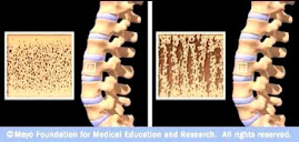
Osteoporotic Bone

Osteoporotic Bone
Source: www.mayoclinic.com

How Strontium Builds Bones

Strontium is a mineral that tends to accumulate in bone. Studies have shown that oral doses of strontium are a safe and effective way to prevent and reverse osteoporosis. Doses of 680 mg per day appear to be optimal. See my "For More Information About Strontium" links section.

Osteoporosis is caused by changes in bone production. In healthy young bones there is a constant cycle of new bone growth and bone removal. With age, more bone is removed and less new bone is produced. The bones become less dense and thus more fragile.

Scientists believe that strontium works in two ways. It may stimulate the replication of pre-osteoblasts, leading to an increase in osteoblasts (cells that build bone). Strontium also directly inhibits the activity of osteoclasts (cells that break down bone). The result is stronger bones.

When taking strontium, be sure to take 1200 mg calcium, 1000 IU vitamin D3, and 500 mg magnesium daily. It is best to take strontium late at night on an empty stomach. Calcium and strontium may compete with each other for absorption if taken together.