Human Skeleton

Human Skeleton

WELCOME TO STRONTIUM FOR BONES BLOG

Have you experienced negative, and even dangerous, side effects from Fosamax (alendronate), Boniva (ibandronate), Actonel (risedronate), Reclast (zoledronic acid), Prolia (denosumab), Forteo (teriparatide), Tymlos (abaloparatide), or other drugs prescribed for osteoporosis? If you have, then rest assured there is a safe, effective treatment for this condition. Strontium, primarily in the form of strontium citrate, is taken orally once a day.

Visitors to my blog can leave comments or ask questions and can remain anonymous, if they wish. Their comments are relayed to my g-mail inbox. Below each post, the number of comments for that post is cited and underlined because it is a link. By clicking on that link below any post, a window opens so that a visitor can leave a comment. Ideally, visitors leave comments on posts most relevant to their comments. All comments to my posts are moderated by me.

Browse the posts and visit the link library of references.


Blog Archive

Showing posts with label bone mineral density. Show all posts

Wednesday, November 11, 2015

Amino Acid Intake and BMD


Jennings et al reported analyses of female identical twins with different amino acid intakes.

Twins with higher intakes of alanine and glycine had higher BMD at the spine than their co-twins, with within-pair differences in spine BMD of 0.012 g/cm2 and 0.014 g/cm2, respectively.

In cross-sectional multivariable analyses of 3,160 females aged 18-79 years, a higher intake of total protein was associated with higher BMD at the spine (quartile Q4 to quartile Q1: 0.017 g/ cm2) and forearm (Q4 to Q1: 0.010 g/cm2.

Intake of alanine, arginine, glutamic acid, leucine, lysine, and proline was associated with higher BMD at the spine and forearm, with the strongest association observed for leucine (Q4 to Q1: 0.024 g/cm2).

When intakes were stratified by source, vegetable or animal, prevalence of osteoporosis or osteopenia was 13% to 19% lower comparing extreme quartiles of vegetable intake for five amino acids (not glutamic acid or proline).

Jennings A, MacGregor A, Spector T, Cassidy A. Amino acid intakes are associated with bone mineral density and prevalence of low bone mass in women: Evidence from discordant monozygotic twins. J Bone Miner Res 2015; doi:10.1002/jbmr.2703.


Saturday, September 12, 2015

Fracture Warnings for Invokana and Invokamet Diabetes Drugs


“The US Food and Drug Administration (FDA) has strengthened its warning for canagliflozin (Invokana, Invokamet, Johnson & Johnson/Janssen) related to the increased risk for bone fractures.”

“The …product label for canagliflozin had already mentioned the risk for bone fractures. Now, based on new confirmatory information from several clinical trials, the FDA has added further warning and precaution information. In the trials, the fractures affected the upper extremities, occurred as early as 12 weeks after starting the drug, and typically arose from minor trauma such as falling from a standing height.”

“The FDA has also added new information to the label about decreased bone mineral density at the hip and lower spine.”

“The FDA is also evaluating the possible risk for bone fractures for other drugs in the sodium glucose cotransporter 2 (SGLT2) inhibitor class, including dapagliflozin (Farxiga, Xigduo XR, AstraZeneca) and empagliflozin (Jardiance, Glyxambi, Synjardy, Lilly/Boehringer Ingelheim), to determine whether additional label changes or studies are needed. The label for Farxiga mentions a small number of cases of fractures in patients with renal impairment; the Jardiance prescribing information does not mention bone effects.”

“…SGLT2 inhibitors increase concentrations of phosphate in serum, probably via increased tubular reabsorption, which has the potential to adversely affect bone.”

“Furthermore…SGLT2 inhibitors increase concentrations of parathyroid hormone (PTH). Sustained increases in PTH concentration enhance bone resorption and increase the risk for bone fractures.”

"Although canagliflozin causes a small increase in mean PTH concentration (7.9%), the standard deviation is large. Thus, a substantial number of patients treated with canagliflozin might have a 50% or greater increase in PTH concentrations — a change that could be clinically significant…"

Simeon I. Taylor, MD, professor of medicine at University of Maryland School of Medicine in Baltimore, said, "Although not proven, I believe that increased risk of bone fracture is likely a class effect. Nevertheless, individual drugs differ with respect to selectivity for SGLT2 vs. SGLT1, and also with respect to where on the dose-response curve the approved dose falls. So, it is certainly possible that the magnitude of the risk could vary among individual SGLT2 inhibitors."


Wednesday, August 27, 2014

Vitamin K2 Controversy


Japanese researchers have published several articles on vitamin K2, such as the following one: “Vitamin K2 (Menatetrenone) Effectively Prevents Fractures and Sustains Lumbar Bone Mineral Density in Osteoporosis” American researchers have been unable to duplicate the results. One study found that “Vitamin K Treatment Reduces Undercarboxylated Osteocalcin but Does Not Alter Bone Turnover, Density, or Geometry in Healthy Postmenopausal North American Women.”

Each study has weaknesses. For example, the Japanese researchers did not use placebo capsules for vitamin K2 in the control group. Therefore, they cannot exclude the possibility that this may have differentially affected the behavior of study participants in the control and treatment groups, although they consider this possibility unlikely. Limitations of the study on North American women include the relatively short (1 yr) study duration and the inclusion of only healthy women. Thus, the exclusion of osteoporotic women and, importantly, the short duration of this study prohibited use of fracture reduction as a study endpoint.

Here are the two abstracts with links to the full articles:

Abstract to Asian Article:
We attempted to investigate whether vitamin K2 (menatetrenone) treatment effectively prevents the incidence of new fractures in osteoporosis. A total of 241 osteoporotic patients were enrolled in a 24-month randomized open label study. The control group (without treatment; n = 121) and the vitamin K2–treated group (n = 120), which received 45 mg/day orally vitamin K2, were followed for lumbar bone mineral density (LBMD; measured by dual-energy X-ray absorptiometry [DXA]) and occurrence of new clinical fractures. Serum level of Glu-osteocalcin (Glu-OC) and menaquinone-4 levels were measured at the end of the follow-up period. Serum level of OC and urinary excretion of deoxypyridinoline (DPD) were measured before and after the treatment. The background data of these two groups were identical. The incidence of clinical fractures during the 2 years of treatment in the control was higher than the vitamin K2–treated group (χ2 = 10.935; p = 0.0273). The percentages of change from the initial value of LBMD at 6, 12, and 24 months after the initiation of the study were −1.8 ± 0.6%, −2.4 ± 0.7%, and −3.3 ± 0.8% for the control group, and 1.4 ± 0.7%, −0.1 ± 0.6%, and −0.5 ± 1.0% for the vitamin K2–treated group, respectively. The changes in LBMD at each time point were significantly different between the control and the treated group (p = 0.0010 for 6 months, p = 0.0153 for 12 months, and p = 0.0339 for 24 months). The serum levels of Glu-OC at the end of the observation period in the control and the treated group were 3.0 ± 0.3 ng/ml and 1.6 ± 0.1 ng/ml, respectively (p < 0.0001), while the serum level of OC measured by the conventional radioimmunoassay (RIA) showed a significant rise (42.4 ± 6.9% from the basal value) in the treated group at 24 months (18.2 ± 6.1% for the controls; p = 0.0081). There was no significant change in urinary DPD excretion in the treated group. These findings suggest that vitamin K2 treatment effectively prevents the occurrence of new fractures, although the vitamin K2–treated group failed to increase in LBMD. Furthermore, vitamin K2 treatment enhances γ-carboxylation of the OC molecule. http://onlinelibrary.wiley.com/doi/10.1359/jbmr.2000.15.3.515/full

Abstract to North American Article
Low vitamin K status is associated with low BMD and increased fracture risk. Additionally, a specific menaquinone, menatetrenone (MK4), may reduce fracture risk. However, whether vitamin K plays a role in the skeletal health of North American women remains unclear. Moreover, various K vitamers (e.g., phylloquinone and MK4) may have differing skeletal effects. The objective of this study was to evaluate the impact of phylloquinone or MK4 treatment on markers of skeletal turnover and BMD in nonosteoporotic, postmenopausal, North American women. In this double-blind, placebo-controlled study, 381 postmenopausal women received phylloquinone (1 mg daily), MK4 (45 mg daily), or placebo for 12 mo. All participants received daily calcium and vitamin D3 supplementation. Serum bone-specific alkaline phosphatase (BSALP) and n-telopeptide of type 1 collagen (NTX) were measured at baseline and 1, 3, 6, and 12 mo. Lumbar spine and proximal femur BMD and proximal femur geometry were measured by DXA at baseline and 6 and 12 mo. At baseline, the three treatment groups did not differ in demographics or study endpoints. Compliance with calcium, phylloquinone, and MK4 treatment was 93%, 93%, and 87%, respectively. Phylloquinone and MK4 treatment reduced serum undercarboxylated osteocalcin but did not alter BSALP or NTX. No effect of phylloquinone or MK4 on lumbar spine or proximal femur BMD or proximal femur geometric parameters was observed. This study does not support a role for vitamin K supplementation in osteoporosis prevention among healthy, postmenopausal, North American women receiving calcium and vitamin D supplementation. http://onlinelibrary.wiley.com/doi/10.1359/jbmr.081254/full


Monday, July 7, 2014

Prevalence and Determinants of Osteoporosis in Patients With Type 1 and Type 2 Diabetes Mellitus


We analyze osteoporosis prevalence and determinants of bone mineral density (BMD) in patients with type 1 and 2 diabetes.  Three hundred and ninety-eight consecutive diabetic patients from a single outpatient clinic received a standardized questionnaire on osteoporosis risk factors and were evaluated for diabetes-related complications, HbA1c levels, and lumbar spine (LS) and femoral neck (FN) BMD. Of these, 139 (71 men, 68 women) type 1 and 243 (115 men, 128 women) type 2 diabetes patients were included in the study. BMD (T-scores and values adjusted for age, BMI and duration of disease) was compared between patient groups and between patients with type 2 diabetes and population-based controls (255 men, 249 women).

For both genders, adjusted BMD was not different between the type 1 and type 2 diabetes groups but was higher in the type 2 group compared with controls. Osteoporosis prevalence (BMD T-score < −2.5 SD) at FN and LS was equivalent in the type 1 and type 2 diabetes groups, but lower in type 2 patients compared with controls (FN: 13.0% vs 21.2%, LS: 6.1% vs 14.9% men; FN: 21.9% vs 32.1%, LS: 9.4% vs 26.9% women). Osteoporosis prevalence was higher at FN-BMD than at LS-BMD. BMD was positively correlated with BMI and negatively correlated with age, but not correlated with diabetes-specific parameters (therapy, HbBA1c, micro- and macrovascular complications) in all subgroups. Fragility fracture prevalence was low (5.2%) and not different between diabetes groups. Fracture patients had lower BMDs compared with those without fractures; however, BMD T-score was above −2.5 SD in most patients.

Our study shows a similar risk of osteoporosis in patients with type 1 diabetes based on low BMD (T-score < −2.5 SD), which was not different from the prevalence in patients with type 2 diabetes despite them being approximately 20 years older. The FN-BMD was particularly decreased; therefore evaluation of osteoporosis risk in younger patients with type 1 diabetes should include both spinal and FN-BMD-measurements.

We found increased LS-BMD and FN-BMD in patients with type 2 diabetes compared with a non-diabetic, population-based control group. There was a trend for lower BMD in diabetic patients (type 1 and 2) with osteoporotic fractures compared with those without fractures; however, the fracture threshold is higher than in non-diabetic populations. Further longitudinal cohort studies are required, focusing on the risk of fractures and changes in bone metabolism in patients with diabetes.

BMD measurements and the evaluation of BMD-independent risk factors for fractures should be included in the routine management of patients with diabetes mellitus because the prediction of osteoporosis solely by clinical diabetes-specific parameters was not possible. The evidence suggests that osteoporosis, and related fractures, is a clinically significant and commonly underestimated problem in patients with type 1 and type 2 diabetes mellitus.
http://www.medscape.com/viewarticle/825846_1

Friday, June 27, 2014

Spinal BMD in Patients with Scoliosis


The purposes of a retrospective study published in the journal, “Spine” were to investigate the validity of bone mineral density measurements with DEXA in patients with adult lumbar scoliosis and to investigate the association between osteoporosis and adult lumbar scoliosis. Osteoporosis and lumbar degenerative scoliosis are phenomena encountered with increased frequency in aging, often concurrently. Scoliotic patients demonstrated increased spinal bone mineral density (BMD) measurements compared with nonscoliotic patients, resulting in discrepancies between hip and spine BMD values. This discrepancy correlated with aging and curve magnitude (up to 30% for curves of 43°). Scoliotic patients demonstrated significantly lower hip BMD values than nonscoliotic. Curve magnitude did not correlate with severity of osteoporosis.

Spinal BMD values are less valuable for monitoring osteoporosis than hip values in scoliotic patients; an increasing discrepancy with age was noted. Scoliotic patients exhibited discordantly high spinal BMD values, despite significant hip osteoporosis. The discrepancy correlated with aging and curve magnitude. Scoliosis was common among the osteoporotic population (9.47%). Lumbar scoliosis is a useful clinical marker for osteoporosis, irrespective of scoliosis history and magnitude. Viable alternatives for osteoporosis evaluation of adult patients with lumbar scoliosis are hip DEXA values, in conjunction with other BMD measurements.


Wandering Skeleton

Wandering Skeleton
Artist: Joel Hoekstra

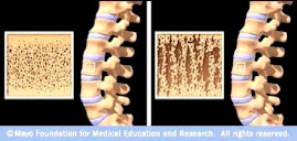
Osteoporotic Bone

Osteoporotic Bone
Source: www.mayoclinic.com

How Strontium Builds Bones

Strontium is a mineral that tends to accumulate in bone. Studies have shown that oral doses of strontium are a safe and effective way to prevent and reverse osteoporosis. Doses of 680 mg per day appear to be optimal. See my "For More Information About Strontium" links section.

Osteoporosis is caused by changes in bone production. In healthy young bones there is a constant cycle of new bone growth and bone removal. With age, more bone is removed and less new bone is produced. The bones become less dense and thus more fragile.

Scientists believe that strontium works in two ways. It may stimulate the replication of pre-osteoblasts, leading to an increase in osteoblasts (cells that build bone). Strontium also directly inhibits the activity of osteoclasts (cells that break down bone). The result is stronger bones.

When taking strontium, be sure to take 1200 mg calcium, 1000 IU vitamin D3, and 500 mg magnesium daily. It is best to take strontium late at night on an empty stomach. Calcium and strontium may compete with each other for absorption if taken together.