Human Skeleton

Human Skeleton

WELCOME TO STRONTIUM FOR BONES BLOG

Have you experienced negative, and even dangerous, side effects from Fosamax (alendronate), Boniva (ibandronate), Actonel (risedronate), Reclast (zoledronic acid), Prolia (denosumab), Forteo (teriparatide), Tymlos (abaloparatide), or other drugs prescribed for osteoporosis? If you have, then rest assured there is a safe, effective treatment for this condition. Strontium, primarily in the form of strontium citrate, is taken orally once a day.

Visitors to my blog can leave comments or ask questions and can remain anonymous, if they wish. Their comments are relayed to my g-mail inbox. Below each post, the number of comments for that post is cited and underlined because it is a link. By clicking on that link below any post, a window opens so that a visitor can leave a comment. Ideally, visitors leave comments on posts most relevant to their comments. All comments to my posts are moderated by me.

Browse the posts and visit the link library of references.


Blog Archive

Showing posts with label bone strontium content. Show all posts

Friday, November 10, 2023

Echolight Scans Are Not Affected by Bone Strontium Content!

We strontium users know that BMD results by DXA scan will be artificially high, especially after years of using strontium salts. Here is good news! Echolight REMS technology is NOT AFFECTED by STRONTIUM. It uses ultrasound instead of ionizing radiation. Echolight is a modern ultrasound device capable of measuring bone at the hip and the spine using Radiofrequency Echographic Multi Spectrometry (REMS).


BMD measured by ultrasound is not affected by the strontium content of bone. An in vitro study concluded that aBMD measured by DXA was prone to overestimation in the presence of strontium, but acoustic parameters measured by quantitative ultrasound (QUS) are independent of strontium concentration.

https://pubmed.ncbi.nlm.nih.gov/29080282/


Another advantage of Echolight scans is that they assess the bone internal micro-architecture.  So, physicians obtain results of bone quantity and quality. 


Yet another advantage of REMS over DXA is that REMS appears to be able to overcome common artifacts such as structural alterations caused by OA (osteoarthritis) at the lumbar spine, which affect the BMD values obtained by DXA

https://www.ncbi.nlm.nih.gov/pmc/articles/PMC10217295/#:~:text=Radiofrequency%20Echographic%20Multi%20Spectrometry%20(REMS,technique%20present%20in%20the%20literaturePMC10217295


Echolight has received 510(k) clearance to sell its EchoS Systems in the USA! Companies must submit a Premarket Notification, also called a PMN or 510(k), to the FDA so that it can review the product and clear it. Once the FDA declares that a new medical device is substantially equivalent to a predicate, it is cleared and can be marketed and sold in the USA. 


Echolight has opened a headquarters in NYC. The scans are not available everywhere. You will have to shop around. One early adopter is Dr. Andrew Bush, M.D., an orthopedic surgeon with a practice that performs Echolight scans in Sanford, North Carolina.

https://www.centralcarolinaortho.com/Blog/374362/EXPLORING-ECHOLIGHT-A-NOVEL-WAY-TO-MONITOR-OSTEOPOROSIS


COPA Healthcare LLC, Dania, FL sells the EchoS/Echolight and Marodyne LiV. The website includes a video that shows the EchoS equipment and how the scan is done. The website makes it easy to leave a message in case you have questions.

https://copahealth.us/echolight/


Monday, September 28, 2020

The Effect of Bone Strontium on BMD for Different Manufacturers' DXA Systems

Osteoporotic patients treated with strontium ranelate show relatively large increases in bone mineral density (BMD) measured by dual-energy X-ray absorptiometry (DXA) due to the replacement of some of the calcium atoms in bone by strontium. A study published by Pors Nielsen and colleagues reported that replacement of 1% of calcium atoms by strontium causes a 10% increase in BMD. 

We refer to the ratio of the percentage increase in BMD to the molar percentage of strontium in bone as the strontium ratio. Theoretically it is expected that the strontium ratio should vary between different manufacturers' DXA equipment depending on the effective photon energy of the device, an effect that arises because of the proximity of the X-ray energies produced by lower energy devices to the strontium K-edge at 16 keV. 

In this study we report theoretical estimates of the strontium ratio for two axial DXA systems and two peripheral DXA devices based on their broad spectrum X-ray emission. The theoretical figures were verified in an experimental study in which the strontium ratio for each device was measured using phantoms containing mixtures of hydroxyapatite and strontium hydrogen-phosphate. 

The theoretical values/experimental results of the strontium ratio were: 

11.0/11.2 for the Hologic Discovery,

9.9/9.9 for the GE-Lunar Prodigy, 

9.1/8.6 for the Demetech Calscan, and 

8.5/6.3 for the Osteometer Dexacare G4. 

The results confirm both theoretically and experimentally that the effect of bone strontium on BMD measurements is different for different DXA systems. In the future it might be possible to exploit this effect to make a non-invasive estimate of average bone strontium content in groups of patients receiving strontium medication for osteoporosis.

https://pubmed.ncbi.nlm.nih.gov/20699129/


Sunday, September 21, 2014

Long-Term Effect of Strontium Ranelate Treatment on BMD


http://onlinelibrary.wiley.com/doi/10.1359/JBMR.050810/full

In the SOTI trial, there were impressive BMD increases in the spine (12.7%) and total hip (8.6%). However, some caution is necessary in interpreting these results because much of this effect is caused by the higher atomic number of strontium (Z = 38) compared with calcium (Z = 20). When BMD is measured by DXA, strontium atoms in bone attenuate X-rays more strongly than calcium atoms. However, when the DXA scanner software calculates BMD from the measured X-ray transmission factors, the increased attenuation caused by bone strontium content (BSC) is interpreted as increased calcium content and will cause an artifactual increase in BMD. After the adjustment for BSC using Eq. 1, the measured BMD increase in the spine at 3 years of 12.7% was corrected to 6.8%.

The correction of the bone mineral content (BMD) changes found in the strontium ranelate trials for bone strontium content (BSC) was based on the following equation:

Adjusted BMD = Measured lumbar spine BMD /1+ 0.061 x BSC iliac crest % Eq. 1

“Although a commendable effort was made in the SOTI trial to correct the BMD data for the atomic number effect of strontium, there is clearly considerable uncertainty about the accuracy of the corrections made. This arises from the small number of subjects in whom iliac crest bone biopsy was performed and the reliance on animal data for the correction factor for inferring BSC in the spine.”

No human studies with strontium ranelate have yet reported how quickly bone strontium is washed out once treatment is stopped. However, on the basis of the ICRP strontium model, we can estimate the likely long-term retention. Figure 2 shows the results of calculations using the ICRP model to predict the long-term changes in BMD after 3 months, 1 year, and 3 years of treatment with strontium ranelate. The calculations make the following assumptions: (1) equal daily intake of strontium during the treatment period; (2) strontium intake ceases at the end of treatment; and (3) no true loss of bone is occurring. On the basis of the ICRP model, much of the strontium present in bone at the end of treatment is likely to still be there a decade later. If strontium ranelate treatment is given for >1 year, this long-term retention in bone is likely to have a significant effect on the interpretation of future BMD measurements.


Tuesday, April 24, 2012

Interpretation of BMD Scans in Patients Stopping Strontium Ranelate Treatment

“The oral administration of strontium ranelate causes a clinically significant overestimation of BMD because of the high attenuation of X-rays by strontium atoms in bone." GM Blake and I. Fogelman “performed a theoretical study to calculate the long-term effect of strontium treatment on BMD and establish the duration of past treatment for which the remaining bone strontium content (BSC) has a negligible effect on BMD.” “Estimates of the strontium BMD artefact after treatment has stopped were performed on the assumption that the BSC effect accounts for 75% of the total measured BMD change at 3 years.” “If 75% of the BMD changes are explained by BSC, in the average patient, 3-year treatment leads to a spine BMD artefact of 11.2%, decreasing to 3.8% 10 years after stopping treatment. The BMD artefacts at the total hip and femoral neck sites are smaller by factors of 0.65 and 0.53, respectively.” “On average, 6-month treatment is required for the spine BMD artefact to exceed 3%, the figure adopted as the maximum BMD change caused by bone strontium that has a negligible effect on scan interpretation.” The researchers concluded that “strontium ranelate treatment lasting for > 6 months can affect BMD measurements for many years afterward.” http://www.ncbi.nlm.nih.gov/pubmed/16939400 J Bone Miner Res. 2006 Sep;21(9):1417-24.

Wandering Skeleton

Wandering Skeleton
Artist: Joel Hoekstra

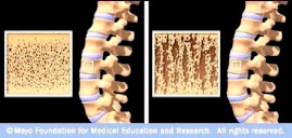
Osteoporotic Bone

Osteoporotic Bone
Source: www.mayoclinic.com

How Strontium Builds Bones

Strontium is a mineral that tends to accumulate in bone. Studies have shown that oral doses of strontium are a safe and effective way to prevent and reverse osteoporosis. Doses of 680 mg per day appear to be optimal. See my "For More Information About Strontium" links section.

Osteoporosis is caused by changes in bone production. In healthy young bones there is a constant cycle of new bone growth and bone removal. With age, more bone is removed and less new bone is produced. The bones become less dense and thus more fragile.

Scientists believe that strontium works in two ways. It may stimulate the replication of pre-osteoblasts, leading to an increase in osteoblasts (cells that build bone). Strontium also directly inhibits the activity of osteoclasts (cells that break down bone). The result is stronger bones.

When taking strontium, be sure to take 1200 mg calcium, 1000 IU vitamin D3, and 500 mg magnesium daily. It is best to take strontium late at night on an empty stomach. Calcium and strontium may compete with each other for absorption if taken together.