Skeleton Pirate

Skeleton Pirate
Artist: LindaB

WELCOME TO STRONTIUM FOR BONES BLOG

Have you experienced negative, and even dangerous, side effects from Fosamax (alendronate), Boniva (ibandronate), Actonel (risedronate), Reclast (zoledronic acid), Prolia (denosumab), Forteo (teriparatide), Tymlos (abaloparatide), or other drugs prescribed for osteoporosis? If you have, then rest assured there is a safe, effective treatment for this condition. Strontium, primarily in the form of strontium citrate, is taken orally once a day.

Visitors to my blog can leave comments or ask questions and can remain anonymous, if they wish. Their comments are relayed to my g-mail inbox. Below each post, the number of comments for that post is cited and underlined because it is a link. By clicking on that link below any post, a window opens so that a visitor can leave a comment. Ideally, visitors leave comments on posts most relevant to their comments. All comments to my posts are moderated by me.

Browse the posts and visit the link library of references.


Blog Archive

Thursday, December 18, 2014

Sequential Treatment with Teriparatide (Forteo) and Strontium Ranelate in a Postmenopausal Woman with Atypical Femoral Fractures after Long-term Bisphosphonate Administration


Abstract
OBJECTIVE:
Despite the existence of numerous case series, no evidenced-based medical management for atypical fractures associated with bisphosphonate (BP) treatment has been established.
DESIGN:
We report the outcome of teriparatide (TRP) administration followed by strontium ranelate (SR) in a woman with a complete and an incomplete contralateral atypical fracture of the femoral diaphysis (AFF) associated with BP treatment. The spontaneous complete AFF was managed with intramedullary nailing, discontinuation of BP and initiation TRP.
RESULTS:
Eleven months later, she suffered a contralateral incomplete AFF. At the completion of the TRP treatment, she had only slight discomfort in the femur with the incomplete AFF. BMD testing revealed increase of 7.61% at the lumbar spine (LS) and 0.8% at the hip. Following TRP, 1-year SR treatment resulted in further BMD increase of 9.2% at the LS and 1.4% in the hip, while she does not report any pain. Bone markers remain within the normal range.
CONCLUSION:
Our case indicates that sequential therapy with TRP and SR in cases of AFF might be a rational treatment option. However, there is a need for additional information concerning the effect of TRP and SR, given alone or sequentially, in these patients in order to incorporate these drugs into the management of AFF.


Study Demonstrates DXA Scan Is Obviously Flawed


Most clinically important vertebral body compression fractures found on abdominal multidetector computed tomography (CT) scans would go unreported if sagittal reconstructions were not routinely evaluated, according to a new study.

"At this point, it is routine to evaluate sagittal reconstructions at abdominal multidetector CT (MDCT), and whenever vertebral fractures are detected, they are reported." However, this has not always been the case, said Travis Lauder, BS, a fourth-year medical student at the University of Wisconsin School of Medicine and Public Health in Madison.

"The studies we reviewed were done before 2007, when sagittal reconstructions were not automatically provided. We showed that without them, we missed a lot of vertebral compression fractures in patients who underwent abdominal multidetector CT," he explained.

Lauder presented the study results at the American Roentgen Ray Society 2013 Annual Meeting in Washington, DC. First results were published online February 28 in Radiology.

Lauder and his team retrospectively analyzed 2041 consecutive patients (mean age, 70.8 years) who had undergone abdominal multidetector CT and dual-energy x-ray absorptiometry (DXA) within 6 months of each other. The images were taken from 2000 to 2007, before sagittal CT reconstructions were routinely obtained.

The researchers used the Genant visual semiquantitative method to review transverse (axial) and retrospective sagittal multidetector CT reconstructions for the presence of moderate to severe fractures of the lower thoracic and lumbar spine. They also reviewed the electronic medical records of these patients to determine whether the fracture was reported at prospective CT interpretation, was known previously, or was subsequently diagnosed.

"At least 1 moderate or severe vertebral body compression fracture was retrospectively identified on CT in 97 patients," the researchers report. The fractures involved 1 level in 67 patients and 2 or more levels in 30 patients. In total, 141 fractures were identified.

In patients with an unreported fracture, vertebral body compression fracture was not recognized clinically in 64% and there was no evidence of osteoporosis on DXA in 48%.

"DXA is still the gold standard," Lauder noted, "but this analysis demonstrated that a lot of these patients have false negatives on DXA, and that the DXA test is obviously flawed."

Incidental findings are a well-known "double-edged sword" in cross-sectional imaging, said Abraham Dachman, MD, professor of radiology and director of the fellowship programs at the University of Chicago in Illinois, who was asked by Medscape Medical News to comment on the study.

"Many findings — some previously known by the referring physician and some not — can be found on CT. It is often difficult for the radiologist to know what findings to report, what findings to ignore, and what incidental findings are sufficiently important to require personal communication with the referring physician," Dr. Dachman said.

However, this study highlights the potential of abdominal CT to detect vertebral body compression fractures, he noted. Coronal and sagittal reconstructions are often done by the technologist who performs the exam using software on the CT scanner console; they do not require dedicated workstations.

"This study did not address how often the observation of vertebral body compression fractures changed patient management because it was retrospective in nature," Dr. Dachman said. "Still, it highlights the potential of CT to reveal information and provides a rationale to radiologists who wish to routinely report this observation."

This article appeared on Medscape under the title, “Vertebral Fractures Go Undetected without Sagittal Views.”
The abstract and full text with CT images for “Unreported Vertebral Body Compression Fractures at Abdominal Multidector CT,” which was published in Radiology  in July 2013, can be read here:
For an excellent description of computed tomography (CT) and multidetector computed tomography (MDCT), see:
For a description of an abdominal CT scan, see:
For information on computed tomography angiography (CTA), see:


Wednesday, December 17, 2014

Strontium Citrate and Strontium Ranelate


Strontium ranelate is prescribed for postmenopausal osteoporosis in the United Kingdom, Australia, much of Europe and Asia, and Mexico. Due to an increased risk of thrombosis and cardiovascular events in patients with a history of cardiovascular problems, some doctors are discouraging their patients from taking strontium ranelate or refusing to prescribe it.

Strontium ranelate is not available in the United States or Canada. However, strontium citrate can be purchased as a supplement from health food stores and online distributors. Is strontium citrate safe and effective?

In 2012, Canadian researchers published the Combination of Micronutrients for Bone (COMB) Study. Here is the abstract:

“Along with other investigations, patients presenting to an environmental health clinic with various chronic conditions were assessed for bone health status. Individuals with compromised bone strength were educated about skeletal health issues and provided with therapeutic options for potential amelioration of their bone health. Patients who declined pharmacotherapy or who previously experienced failure of drug treatment were offered other options including supplemental micronutrients identified in the medical literature as sometimes having a positive impact on bone mineral density (BMD). After 12 months of consecutive supplemental micronutrient therapy with a combination that included vitamin D3, vitamin K2, strontium, magnesium and docosahexaenoic acid (DHA), repeat bone densitometry was performed. The results were analyzed in a group of compliant patients and demonstrate improved BMD in patients classified with normal, osteopenic and osteoporotic bone density. According to the results, this combined micronutrient supplementation regimen appears to be at least as effective as bisphosphonates or strontium ranelate in raising BMD levels in hip, spine, and femoral neck sites. No fractures occurred in the group taking the micronutrient protocol. This micronutrient regimen also appears to show efficacy in individuals where bisphosphonate therapy was previously unsuccessful in maintaining or raising BMD. Prospective clinical trials are required to confirm efficacy.”

Here is the COMB protocol for bone health:
(1) Docosahexaenoic acid or DHA (from Purified Fish Oil): 250mg/day          
(2) Vitamin D3: 2000IU/day         
(3) Vitamin K2 (non-synthetic MK7 form): 100ug/day        
(4) Strontium citrate: 680mg/day (strontium)       
(5) Elemental magnesium: 25mg/day      
(6) Dietary sources of calcium recommended        
(7) Daily impact exercising encouraged    

The following two paragraphs are quotes from the paper:
“Studies to date have predominantly focused on strontium ranelate rather than the readily available strontium citrate supplement as used in this study. The results of this study, however, demonstrate that the micronutrient combination including strontium citrate is at least as effective in BMD change as strontium ranelate with suggestion of preferred efficacy of the former therapy at improving femoral neck outcomes. Furthermore, the ranelic acid salt is a purely synthetic molecular compound, while citrate is naturally occurring. It appears to be the strontium portion of the molecules which exerts most or all of the positive effect on bone. When consuming the strontium ranelate, for example, the compound splits into two strontium ions and one molecule of ranelic acid, with each absorbed separately. There is little evidence that the ranelic acid portion of the strontium ranelate compound contributes to the effect of strontium on skeletal tissue, and of the small amount of ranelic acid that is absorbed into the body, almost all is excreted within a week without ever being metabolized. All forms of strontium have bioavailabilities in the 25–30% range, but gastric tolerance appears to be better with the ranelate and citrate forms.”

“With the mounting concern about the safety profile of some standard medical interventions for bone compromise, strontium is very well tolerated and has shown remarkably little in the way of side effects or long-term adverse sequelae. An increased risk of thrombosis has been noted with strontium ranelate, an effect not reported (to our knowledge) with strontium citrate [16].”

16. S. J. Genuis and G. K. Schwalfenberg, “Picking a bone with contemporary osteoporosis management: nutrient strategies to enhance skeletal integrity,” Clinical Nutrition, vol. 26, no. 2, pp. 193–207, 2007.
The abstract to reference 16:
 http://www.clinicalnutritionjournal.com/article/S0261-5614%2806%2900151-8/abstract
The entire article (reference 16) in PDF form:
 http://www.uio.no/studier/emner/medisin/nutri/ERN3120/v12/ERN3120-Unit3-Student/02%20Osteoporosis%20and%20Ostaomalacia/Osteoporose-ern%C3%A6ring-03.pdf

The entire COMB Study article can be read here:


Monday, December 15, 2014

Fixed Risk Factors for Osteoporosis


Fixed risk factors determine whether an individual is at heightened risk of osteoporosis. Also, unlike modifiable risks, they are factors which we can’t change, including age, gender and family history.
In addition, people may have secondary risk factors. These include disorders and medications that weaken bone and affect balance (heightening the risk of fracture due to falling). Read more about Secondary Osteoporosis.

Low bone mineral density, one of the most important indicators that a person is at risk of a fracture, is considered both fixed and modifiable since it is determined by a wide range of factors, including family history, age and lifestyle factors.

Although fixed risk factors for osteoporosis cannot be changed, people need to be aware of these risks so that they can take steps to reduce bone mineral loss as early as possible. These risks include:
Age
The majority of hip fractures (90%) occur in people aged 50 and older. This is partly due to reduced bone mineral density as we age. But age can also be a risk factor independent of bone mineral density. In other words, even older adults with normal bone mineral density are more likely to suffer a fracture than younger people1.
Female gender
Women, particularly post-menopausal women, are more susceptible to bone loss than men, because their bodies produce less estrogen. This hormone is an important component in bone formation. Women are more likely to sustain an osteoporotic fracture than men. Lifetime risk of any fracture ranges between 40-50% in women, compared to 13-22% in men.
Family history
A parental history of fracture (particularly a family history of hip fracture) is associated with an increased risk of fracture that is independent of bone mineral density2.
Previous fracture
A previous fracture increases the risk of any fracture by 86%, compared with people without a prior fracture. Both men and women are almost twice (1.86 times) as likely to have a second fracture compared to people who are fracture free3.
Ethnicity
Studies have found osteoporosis is more common in Caucasian and Asian populations, and the incidence of osteoporosis and fractures of the hip and spine is lower in black than in white people.
Menopause or hysterectomy
Hysterectomy, if accompanied by removal of the ovaries, may also increase the risk for osteoporosis because of estrogen loss. Post-menopausal women, and those who have had their ovaries removed, must be particularly vigilant about their bone health.
Long term glucocorticoid therapy
Long-term corticosteroids use is a very common cause of secondary osteoporosis and is associated with an increased risk of fracture4.
Rheumatoid arthritis
Rheumatoid arthritis and diseases of the endocrine system can take a heavy toll on bones. Hyperparathyroidism, for example, results in elevated levels of parathyroid hormone, which signals bone cells to release calcium from bone into the blood.
Primary or secondary hypogonadism in men
Like estrogen deficiency in women (which is observed in case of primary or secondary amenorrhea and premature menopause), androgen deficiency in men (primary or secondary hypogonadism) increases the risk of fracture.

At any age, acute hypogonadism, such as that resulting from orchidectomy for prostate cancer, accelerates bone loss to a similar rate as seen in menopausal women. The bone loss following orchidectomy is rapid for several years, and then reverts to the gradual loss that normally occurs with aging.
Secondary Risk Factors
Secondary risk factors are less prevalent but they can have a significant impact on bone health and fracture incidence. These risk factors include other diseases that directly or indirectly affect bone remodeling and conditions that affect mobility and balance, which can contribute to the increased risk of falling and sustaining a fracture.
Disorders that affect the skeleton:
  • Asthma
  • Nutritional/gastrointestinal problems (e.g. Crohn’s or celiac disease)
  • Rheumatoid arthritis
  • Hematological disorders/malignancy
  • Some inherited disorders
  • Hypogonadal states (e.g. Turner syndrome/Kleinfelter syndrome, amenorrhea)
  • Endocrine disorders (e.g. Cushing’s syndrome, hyperparathyroidism, diabetes)
  • Immobility
Medical treatments affecting bone health:

Some medications may have side effects that directly weaken bone or increase the risk of fracture due to fall or trauma. Patients taking any of the following medications should consult with their doctor about increased risk to bone health.
  • Glucocorticosteroids
  • Certain immunosuppressant (calmodulin/calcineurine phosphatase inhibitors)
  • Thyroid hormone treatment (L-Thyroxine)
  • Certain steroid hormones (medroxyprogesterone acetate, leutenising hormone releasing hormone agonists)
  • Aromatase inhibitors
  • Certain antipsychotics
  • Certain anticonvulsants
  • Certain antiepileptic drugs
  • Lithium
  • Methotrexate
  • Antacids
  • Proton pump inhibitors 
References
1. Kanis JA, Johnell O, Odén A, Dawson A, De LAet C, Jonsson B. Ten year probabilities of osteoporotic fractures according to BMD and diagnosis thresholds. Osteoporosis Int 2001; 12:989-95
2. Kanis JA, Johansson H, Odén A, Johnell O, De LAet C, Eisman JA, McCloskey EV, Mellström D, Melton LJ III, Pols HA, Reeve J, Silman AJ, Tenenhouse A. A family history of fracture and fracture risk: a meta-analysis. Bone 2004; 35:1029-37
3. Kanis JA, De LAet C, Delmas P, Garnero P, Johansson H, Johnell O, Kriger H, McCloskey EV, Mellstrom D, Melton LJ III, Odén A, Pols H, Reeve J, Silman A, tenehouse A. A meta-analysis of previous fracture and fracture risk. Bone 2004; 35: 375-82
4. Kanis J A, Johansson H, Odén A, Johnell O, De Laet C, Melton LJ III, Tenenhouse A, Reeve J, Silman AJ, Pols H, Eisman JA, McCliskey EV, Mellström D. A meta-analysis of prior corticosteroid use and fracture risk. J Bone and Miner Res 2004;19: 893-99

Wandering Skeleton

Wandering Skeleton
Artist: Joel Hoekstra

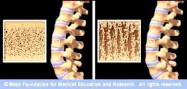
Osteoporotic Bone

Osteoporotic Bone
Source: www.mayoclinic.com

How Strontium Builds Bones

Strontium is a mineral that tends to accumulate in bone. Studies have shown that oral doses of strontium are a safe and effective way to prevent and reverse osteoporosis. Doses of 680 mg per day appear to be optimal. See my "For More Information About Strontium" links section.

Osteoporosis is caused by changes in bone production. In healthy young bones there is a constant cycle of new bone growth and bone removal. With age, more bone is removed and less new bone is produced. The bones become less dense and thus more fragile.

Scientists believe that strontium works in two ways. It may stimulate the replication of pre-osteoblasts, leading to an increase in osteoblasts (cells that build bone). Strontium also directly inhibits the activity of osteoclasts (cells that break down bone). The result is stronger bones.

When taking strontium, be sure to take 1200 mg calcium, 1000 IU vitamin D3, and 500 mg magnesium daily. It is best to take strontium late at night on an empty stomach. Calcium and strontium may compete with each other for absorption if taken together.