Human Skeleton

Human Skeleton

WELCOME TO STRONTIUM FOR BONES BLOG

Have you experienced negative, and even dangerous, side effects from Fosamax (alendronate), Boniva (ibandronate), Actonel (risedronate), Reclast (zoledronic acid), Prolia (denosumab), Forteo (teriparatide), Tymlos (abaloparatide), or other drugs prescribed for osteoporosis? If you have, then rest assured there is a safe, effective treatment for this condition. Strontium, primarily in the form of strontium citrate, is taken orally once a day.

Visitors to my blog can leave comments or ask questions and can remain anonymous, if they wish. Their comments are relayed to my g-mail inbox. Below each post, the number of comments for that post is cited and underlined because it is a link. By clicking on that link below any post, a window opens so that a visitor can leave a comment. Ideally, visitors leave comments on posts most relevant to their comments. All comments to my posts are moderated by me.

Browse the posts and visit the link library of references.


Blog Archive

Showing posts with label vertebral fractures. Show all posts

Thursday, December 18, 2014

Study Demonstrates DXA Scan Is Obviously Flawed


Most clinically important vertebral body compression fractures found on abdominal multidetector computed tomography (CT) scans would go unreported if sagittal reconstructions were not routinely evaluated, according to a new study.

"At this point, it is routine to evaluate sagittal reconstructions at abdominal multidetector CT (MDCT), and whenever vertebral fractures are detected, they are reported." However, this has not always been the case, said Travis Lauder, BS, a fourth-year medical student at the University of Wisconsin School of Medicine and Public Health in Madison.

"The studies we reviewed were done before 2007, when sagittal reconstructions were not automatically provided. We showed that without them, we missed a lot of vertebral compression fractures in patients who underwent abdominal multidetector CT," he explained.

Lauder presented the study results at the American Roentgen Ray Society 2013 Annual Meeting in Washington, DC. First results were published online February 28 in Radiology.

Lauder and his team retrospectively analyzed 2041 consecutive patients (mean age, 70.8 years) who had undergone abdominal multidetector CT and dual-energy x-ray absorptiometry (DXA) within 6 months of each other. The images were taken from 2000 to 2007, before sagittal CT reconstructions were routinely obtained.

The researchers used the Genant visual semiquantitative method to review transverse (axial) and retrospective sagittal multidetector CT reconstructions for the presence of moderate to severe fractures of the lower thoracic and lumbar spine. They also reviewed the electronic medical records of these patients to determine whether the fracture was reported at prospective CT interpretation, was known previously, or was subsequently diagnosed.

"At least 1 moderate or severe vertebral body compression fracture was retrospectively identified on CT in 97 patients," the researchers report. The fractures involved 1 level in 67 patients and 2 or more levels in 30 patients. In total, 141 fractures were identified.

In patients with an unreported fracture, vertebral body compression fracture was not recognized clinically in 64% and there was no evidence of osteoporosis on DXA in 48%.

"DXA is still the gold standard," Lauder noted, "but this analysis demonstrated that a lot of these patients have false negatives on DXA, and that the DXA test is obviously flawed."

Incidental findings are a well-known "double-edged sword" in cross-sectional imaging, said Abraham Dachman, MD, professor of radiology and director of the fellowship programs at the University of Chicago in Illinois, who was asked by Medscape Medical News to comment on the study.

"Many findings — some previously known by the referring physician and some not — can be found on CT. It is often difficult for the radiologist to know what findings to report, what findings to ignore, and what incidental findings are sufficiently important to require personal communication with the referring physician," Dr. Dachman said.

However, this study highlights the potential of abdominal CT to detect vertebral body compression fractures, he noted. Coronal and sagittal reconstructions are often done by the technologist who performs the exam using software on the CT scanner console; they do not require dedicated workstations.

"This study did not address how often the observation of vertebral body compression fractures changed patient management because it was retrospective in nature," Dr. Dachman said. "Still, it highlights the potential of CT to reveal information and provides a rationale to radiologists who wish to routinely report this observation."

This article appeared on Medscape under the title, “Vertebral Fractures Go Undetected without Sagittal Views.”
The abstract and full text with CT images for “Unreported Vertebral Body Compression Fractures at Abdominal Multidector CT,” which was published in Radiology  in July 2013, can be read here:
For an excellent description of computed tomography (CT) and multidetector computed tomography (MDCT), see:
For a description of an abdominal CT scan, see:
For information on computed tomography angiography (CTA), see:


Thursday, December 11, 2014

FRAX Identifies Women with Prevalent Asymptomatic Vertebral Fractures


Abstract

Background
A Moroccan model for the FRAX tool to determine the absolute risk of osteoporotic fracture at 10 years has been established recently. The study aimed to assess the discriminative capacity of FRAX in identifying women with prevalent asymptomatic vertebral fractures (VFs).

Methods
We enrolled in this cross-sectional study 908 post-menopausal women with a mean age of 60.9 years ±7.7 (50 to 91) with no prior known diagnosis of osteoporosis. Subjects were recruited from asymptomatic women selected from the general population. Lateral VFA (vertebral fracture assessment) images and scans of the lumbar spine and proximal femur were obtained using a GE Healthcare Lunar Prodigy densitometer. VFs were defined using a combination of Genantsemiquantitative (SQ) approach and morphometry. We calculated the absolute risk of major fracture and hip fracture with and without bone mineral density (BMD)using the FRAX website.The overall discriminative value of the different risk scores was assessed by calculating the areas under the ROC curve (AUC).

Results
VFA images showed that 179 of the participants (19.7%) had at least one grade 2/3 VF. The group of women with VFs had a statistically significant higher FRAX scores for major and hip fractures with and without BMD, and lower weight, height, and lumbar spine and hip BMD and T-scores than those without a VFA-identified VF. The AUC ROC of FRAX for major fracture without BMD was 0.757 (CI 95%; 0.718-0.797) and 0.736 (CI 95%; 0.695-0.777) with BMD, being 0.756 (CI 95%; 0.716-0.796) and 0.747 (CI 95%; 0.709-0.785), respectively for FRAX hip fracture without and with BMD. The AUC ROC of lumbar spine T-score and femoral neck T-score were 0.660 (CI 95%; 0.611-0.708) and 0.707 (CI 95%; 0.664-0.751) respectively.

Conclusion
In asymptomatic post-menopausal women, the FRAX risk for major fracture without BMD had a better discriminative capacity in identifying the women with prevalent VFs than lumbar spine and femoral neck T-scores suggesting its usefulness in identifying women in whom VFA could be indicated. 

To read the full research article by Abdellah El Maghraoui, Siham Sadni, Nabil Jbili, Asmaa Rezqi, Aziza Mounach and Imad Ghozlani click on this link:

 http://www.biomedcentral.com/1471-2474/15/365


Thursday, August 7, 2014

Effect of Strontium Ranelate on the Risk of Osteoporotic Fractures


Summary
The aim of the present study was to determine the efficacy of strontium ranelate as a function of baseline fracture risk. Treatment with strontium ranelate was associated with a significant 31% decrease in all clinical osteoporotic fractures (vertebral fractures included). Hazard ratios for the effect of strontium ranelate on the fracture outcome did not change significantly with increasing fracture probability.

Introduction
 Two previous studies have suggested that the efficacy of intervention may be greater in the segment of the population at highest fracture risk as assessed by the FRAX® algorithms. The aim of the present study was to determine whether the anti-fracture efficacy of strontium ranelate was dependent of the level of fracture risk.

Methods
The primary data of the two phase III studies (SOTI and TROPOS) of the effects of strontium ranelate in postmenopausal osteoporosis were combined. Country-specific probabilities were computed using the FRAX® tool (version 2.0). The primary outcome variable comprised all clinical osteoporotic fractures (including clinical vertebral fractures). Interactions between fracture probability and efficacy were explored by Poisson regression.

Results
The 10-year probability of major osteoporotic fractures (with BMD) ranged from 2.5% to 90.8%. FRAX®-based hip fracture probabilities ranged from 0.1% to 90.3%. The incidence of clinical osteoporotic fractures (vertebral fractures excluded) and morphometric vertebral fractures increased with increasing baseline fracture probabilities. Treatment with strontium ranelate was associated with a 31% (95% CI=20–39%) decrease in osteoporotic clinical fractures and a 40% decrease in vertebral fractures assessed by semiquantitative morphometry (95% CI=31–48%) Hazard ratios for the effect of strontium ranelate on the fracture outcomes did not change significantly with increasing fracture probability.

Conclusion
Strontium ranelate significantly decreased the risk of osteoporotic clinical fractures, non vertebral fractures and morphometric vertebral fractures in women. Overall, the efficacy of strontium ranelate was not dependent of the level of fracture risk assessed by FRAX.

 
http://link.springer.com/article/10.1007/s00198-010-1474-0#page-1

Wandering Skeleton

Wandering Skeleton
Artist: Joel Hoekstra

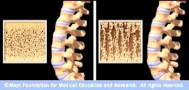
Osteoporotic Bone

Osteoporotic Bone
Source: www.mayoclinic.com

How Strontium Builds Bones

Strontium is a mineral that tends to accumulate in bone. Studies have shown that oral doses of strontium are a safe and effective way to prevent and reverse osteoporosis. Doses of 680 mg per day appear to be optimal. See my "For More Information About Strontium" links section.

Osteoporosis is caused by changes in bone production. In healthy young bones there is a constant cycle of new bone growth and bone removal. With age, more bone is removed and less new bone is produced. The bones become less dense and thus more fragile.

Scientists believe that strontium works in two ways. It may stimulate the replication of pre-osteoblasts, leading to an increase in osteoblasts (cells that build bone). Strontium also directly inhibits the activity of osteoclasts (cells that break down bone). The result is stronger bones.

When taking strontium, be sure to take 1200 mg calcium, 1000 IU vitamin D3, and 500 mg magnesium daily. It is best to take strontium late at night on an empty stomach. Calcium and strontium may compete with each other for absorption if taken together.