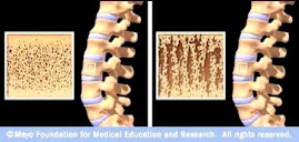
Dual x-ray absorptiometry (DXA) is
currently the standard for assessing bone mineral density (BMD) and has been
correlated with fracture risk and treatment efficacy. DXA includes the
posterior elements of the spine, and therefore
may be inaccurate or not possible in cases of severe spinal degeneration,
scoliosis, or following lumbar surgery. T-score evaluations are somewhat
limited in clinical utility, as the majority of patients who sustain fragility
fractures are not in the osteoporotic range.
Assessing local bone quality on CT scans with Hounsfield
unit (HU) quantification is being used with increasing frequency. Correlations
between HU and bone mineral density have been established, and normative data
have been defined throughout the spine. Recent investigations have explored the
utility of HU values in assessing fracture risk, implant stability, and spinal
fusion success. The information provided by a simple HU measurement can alert
the treating physician to decreased bone quality, which can be useful in both
medically and surgically managing these patients.
The purpose of this paper is to
review the reliability and validity of the techniques used to estimate bone
health using CT scans with Hounsfield unit (HU) quantification. Such scans can
be used to identify patients at risk for osteoporosis, and these values could
be used for surgical planning in cases of trauma, degeneration, and deformity.
There was good correlation of HU value to DXA for both BMD and T-score.


No comments:
Post a Comment